Poietis, a French biotechnology company, has announced the granting of a third patent for its laser-assisted 3D bioprinting method.
“We were the first to explore the industrial potential of laser-assisted bioprinting technology in various applications,” stated Fabien Guillemot, President and CEO of Poietis.
“The granting of this series of European patents further supports Poietis’ position in the field of 3D bioprinting.”
Laser-assisted 3D bioprinting
With five years of experience in the medical field, Poietis’ 3D bioprinting technology presents a novel process of developing realistic self-organizing cell structures. “Unlike conventional approaches to tissue engineering or extrusion bioprinting, laser-assisted bioprinting allows cells to be positioned in three dimensions with micrometric resolution and precision,” Poeitis states.
Its technology is being implemented in cosmetics to fabricate skin models, through a partnership with BASF. Poietis has also partnered with Servier Laboratories, a privately owned global pharmaceutical company, to create a liver toxicity detection model capable of identifying drug-induced lesions.
Most recently, the company has introduced a 4D bioprinting method in which tissue self-organization is programmed by designing the initial tissue architecture in CAD. This improves the functionality of the 3D bioprinted tissues.
4D bioprinted skin equivalent models. Photo via Poietis.
The new Poietis patent
Previously delivered in France, the latest patent issued by the European Patent Office, Patent No. EP3233499, is entitled “Laser printing method and device for implementing said method.” It follows the 2017 Patent No. EP2542659 entitled “Bioprinting Station, assembly comprising such
bioprinting station and bioprinting method” which relates to the adaptation of the printing patterns on substrates.
Moreover, in June 2018, Poietis had been issued Patent No. EP3234102 entitled “Method for laser printing biological components and device for implementing said method”, which covers the laser-assisted bioprinting and multimodal bioprinting processes in Europe.
“The grant of these patents strengthens Poietis’ intellectual property asset,” explained Bruno Brisson, General Manager and VP Business Development at Poietis. “It is also additional protection of important technological bricks that are integrated into the NGB-R, next-gen bioprinting systems that we commercialize now.”
For the latest medical additive manufacturing news, subscribe to our3D printing newsletter and follow usFacebook andTwitter. Visit our3D Printing Jobs board to find out more about opportunities in additive manufacturing.
Featured image shows the NGB-R 3D bioprinter manufactured a 16 layers structure. Photo via Poietis.
Shapeways, a 3D printing marketplace and service bureau, has added Nylon PA11 to its materials portfolio to enable the creation of 3D printed orthotics and prosthetics. As a result of an ongoing partnership with leading 3D printer OEM EOS, Shapeways’ offering of Nylon PA11 has been undertaken to increase 3D printing’s accessibility for medical professionals.
“Expanding our reach to the medical industry to make life-changing enhancements available is something we could have only imagined when we first opened our doors,” said Greg Kress, CEO of Shapeways.
“We look forward to working with the medical industry and creating products [that] make someone’s life better. This is what the 3D printing industry has been working towards and making it available to businesses of all sizes is a real dream.”
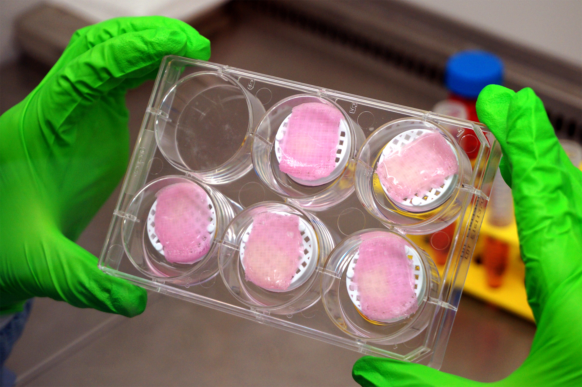
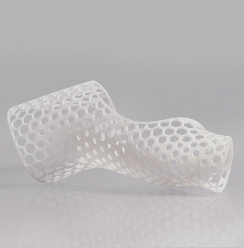
A 3D printed cast using Nylon PA11. Photo via Shapeways.
Shapeways and EOS
Shapeway was founded in 2007 and, at the time, EOS became one of the first additive manufacturing names to become a partner of the site. Headquartered in New York City, Shapeways facilities across the U.S. and Europe house multiple EOS 3D printers.
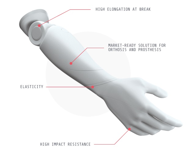
The EOS P 396 3D printer in particular, is enabling the production of PA11 parts at Shapeways. With high impact resistance and low water absorption PA11 is well suited to external medical applications. As such, Shapeways expects to be producing the links of prosthesis, braces, insoles and other supportive devices with the material.
Glynn Fletcher, President, EOS North America, comments, “PA11 has the potential to transform how the medical industry can use 3D printing to improve patient outcomes. It has the additional benefit of lowering the negative, ecological effects that are generally associated with petroleum-based plastics.”
A 3D prosthetic arm using Nylon PA11. Image via Shapeways.
Shapeways expanding capabilities
In 2018, Shapeways was granted a further $30 million in funding to expand its services. In recent months, the company has also been broadening its 3D printing capabilities first in a collaboration with Stratasys and, following this, a partnership with Silicon Valley’s Carbon, giving Shapeways customers access to Digital Light Synthesis (DLS) technology.
Of the most recent PA11 addition, Fletcher concludes, “Collaborating with the industry visionaries at Shapeways has always felt like a privilege. Working together to combine their expertise, our technology with an eco-friendly polymer breakthrough seems like an appropriate extension to our complementary pioneering attitude.”
“We are proud to be part of the venture and look forward to seeing how far the technology, material, and collaboration can take us.”
This method, presented in the journal Microsystems and Nanoengineering, was created to produce lattice scaffolds that enable precise control over its environment as well as cultured cells with particular characteristics.
“If you take cells and put them on a conventional 3D printed surface, it’s like a 2D surface to them, because the cells themselves are so much smaller,” explained Filippos Tourlomousis, a postdoctoral associate at MIT’s Center for Bits and Atoms, and first author of the research.
“But in a mesh-like structure printed using the electro-writing method, the structure is at the same size scale as the cells themselves, and so their sizes and shapes and the way they form adhesions to the material can be controlled by adjusting the porous microarchitecture of the printed lattice structure.”
3D bioprinting and melt electro writing
Tourlomousis and six others at MIT and the Stevens Institute of Technology in New Jersey sought out to accurately produce and tune cells in order to observe and control cell phenotypes; this is due to the need for tighter control over cellular function. According to the researchers, this is a major roadblock for getting tissue engineering products to the clinic.
“Any steps to tighten specifications on the scaffold, and thereby also tighten the variance in cell phenotype, are much needed by this industry,” added Tourlomousis.
“While ordinary 3D printing produces filaments as fine as 150 microns (millionths of a meter), it’s possible to get fibers down to widths of 10 microns by adding a strong electric field between the nozzle extruding the fiber and the stage on which the structure is being printed.” This method is known as melt electrowriting.
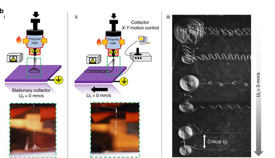
b) Direct melt electrowriting (MEW) by (i) 3D conical fiber structures are obtained by the layered deposition of fibers in circular patterns. (ii) The movement of the grounded collector plate at prescribed translational stage speeds. (iii) Micrograph depicting various fiber topographies that are obtained by tuning the translational stage speed. Image via MIT.
The study explains that cells form proteins known as focal adhesions at the places where they attach themselves to the structure. “Focal adhesions are the way the cell communicates with the external environment. These proteins have measurable features across the cell body allowing us to do metrology. We quantify these features and use them to model and classify quite precisely individual cell shapes.”
Tourlomousis stated, “It is widely known that cell shape governs cell function and this work suggests a shape-driven pathway for engineering and quantifying cell responses with great precision and with great reproducibility.”
The researchers believe this method could be used to 3D print metamaterials that can produce rare optical or electronic properties.
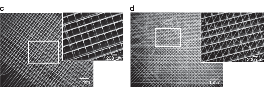
c) 3D woven fibrous mesh with “0–90°” pore microarchitecture fabricated with direct melt electrowriting (MEW). The sample is designated MEW|0–90°. d) 3D woven fibrous mesh with “0–45°” pore microarchitecture. The sample is designated as MEW|0–45°. Image via MIT.
Looking for a career in additive manufacturing? Visit 3D Printing Jobs for a selection of roles in the industry.
Featured image shows Microfilaments made using a new 3D printing method, shown in gray in this illustration, form a structure that cells, shown in color, can adhere to. The shapes formed by the filaments determine the very uniform shapes of cells. Image via Eli Gershenfeld/MIT.
Engineers from McMaster University in Hamilton, Canada, have developed a 3D bioprinting method using magnets to rapidly produce cell clusters.
With formulated bioinks comprised of human breast cancer cells and magnetic salt hydrate, also known as Gd-DTPA, a magnetic field is applied, displacing the cells to a fixed area which “seeds” the formation of a 3D cell cluster.
In a study published in Research, a Science partner journal, the engineers use this method to 3D print cancer tumors within six hours. This process is designed to create an alternative laboratory environment mimicking conditions inside the body for regenerative medicine research – reducing the need for animal testing.
“We have developed a technical solution to overcome the current biological limitations and has the potential to accelerate tissue engineering and regenerative medicine technology,” said Sarah Mishriki, a Ph.D. Candidate at the School of Biomedical Engineering and lead author of the study.
“The ability to rapidly manipulate cells in a safe, controllable and non-contact manner allows us to create the unique cell scapes and microarchitectures of human tissues without the use of scaffolding.”
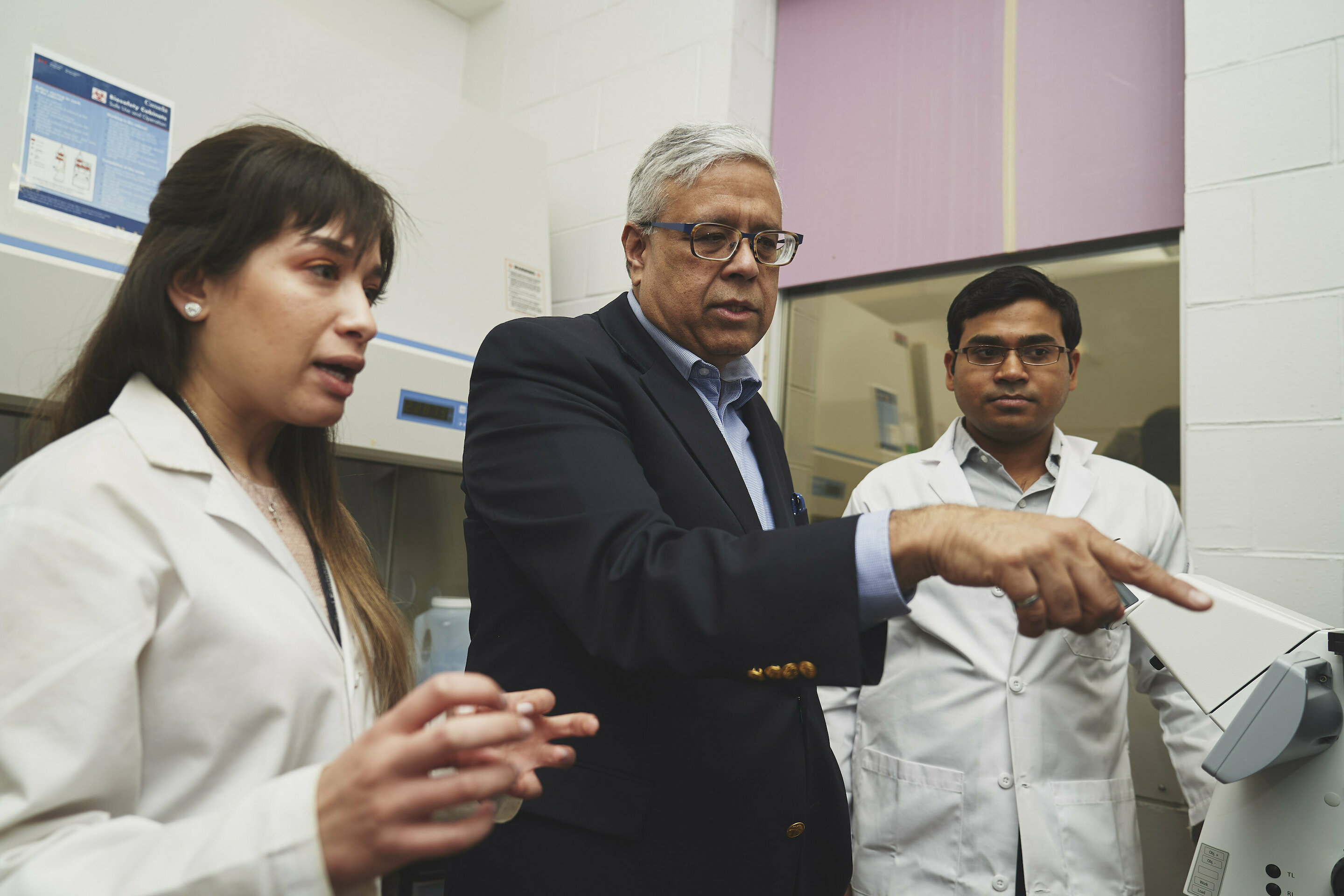
Ishwar Puri, center, with his research team, Sarah Mishriki, PhD candidate in the School of Biomedical Engineering and lead author, and Rakesh Sahu, a research associate. Photo via Jin Lee, McMaster University, Faculty of Engineering.
Accelerating regenerative research
Typically, 2D cell cultures grown in Petri dishes models are less likely to accurately interact than those produced in a 3D environment. 2D models also consist of animal cells which enable drug testing and the study of disease progression.
Ishwar Puri, a professor of mechanical engineering and biomedical engineering, led the McMaster team to develop a new method to rapidly print 3D cell clusters that can better mimic conditions inside the body without the use of animal cells.
This was done using magnetic fields and carefully arranged magnets, which controlled the volume of the cancer cell-laden bioinks. Using this method, the team was able to 3D print cancer tumors. Rakesh Sahu, a research associate, added:
“This magnetic method of producing 3D cell clusters takes us closer to rapidly and economically creating more complex models of biological tissues, speeding discovery in academic labs and technology solutions for industry.”
The researchers hope this method will lead to faster drugs tests using artificial human tumors which can foster breakthroughs in the treatment of cancer and tissue engineering.
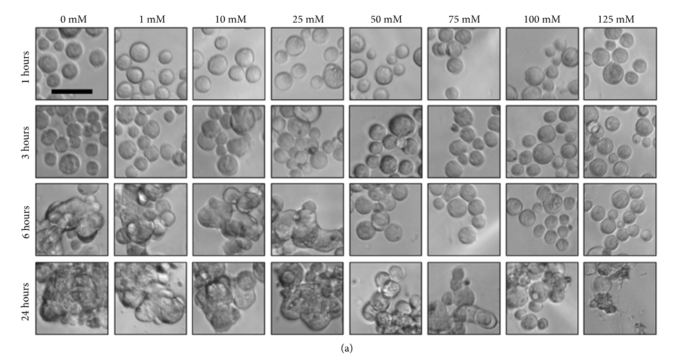
Effect of Gd-DTPA on cell morphology. The breast cancer cells morphologies accumulate malignant plasma cells (mM) in a range from 0, 1, 10, 25, 50, 75, 100, and 125 within 24 hours. Image via McMaster University.
Looking for a career in additive manufacturing? Visit 3D Printing Jobs for a selection of roles in the industry.
Featured image shows Ishwar Puri, center, with his research team, Sarah Mishriki, Ph.D. candidate in the School of Biomedical Engineering and lead author, and Rakesh Sahu, a research associate. Photo via Jin Lee, McMaster University, Faculty of Engineering.
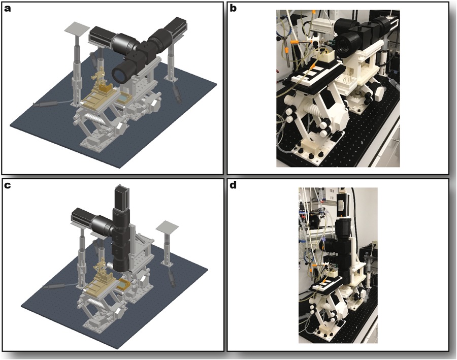
In a study published in Scientific Reports, the researchers sought to create a customizable and cost-effective optical mapping system to understand the relationship between the electrical and mechanical activity of intact hearts.
Such optical mapping systems use the process of perfusion with fluorescent dyes through tissue. “A number of intracellular parameter changes could be tracked this way using high-speed cameras,” the research states; this leads to the mapping of cardiovascular activity.
“The high cost of the equipment and the technical challenges of monitoring multiple parameters of the sample at once and processing the associated signals prevent more widespread use of optical mapping in the biological community.”
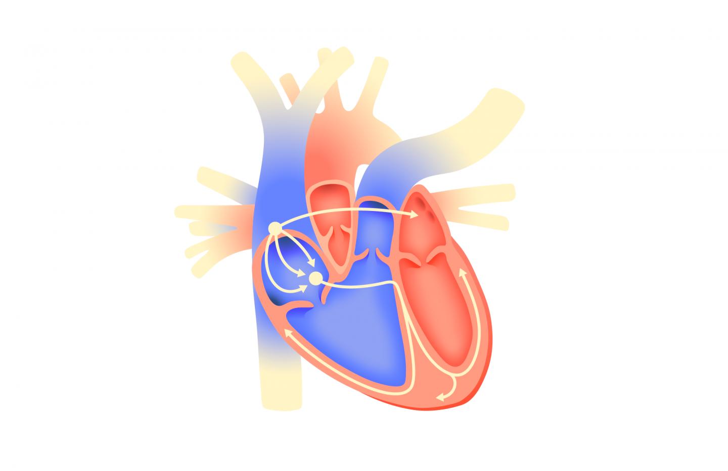
An illustration depicting an electrical excitation initiated in the right atrium spreading through the cardiac conduction system. Image via tsarcyanide/MIPT.
3D printed optical mapping
According to the research team, the mapping process uses cardiac excitation; this primarily involves electrical excitation and the variation in calcium concentration which affect blood levels in the heart. “Normally, excitation is initiated by a group of cells in the right atrium, called the sinoatrial node, and spreads through the cardiac conduction system to the atria and ventricles.”
“Abnormalities in propagation, known as arrhythmias, are a leading cause of mortality in Russia and other developed countries.”
Using the Stratasys Fortus 250mc 3D printer, open-source optical components excluding cameras, lenses, and pumps, were additively manufactured. The researchers have calculated that this system, which operated using Matlab-based multi-signal processing software, could save other researchers up to $20,000 when compared with commercially available products.
“Studies using multiparametric mapping are still uncommon,” explained Roman Syunyaev, a researcher at MIPT’s Human Physiology LabSyunyaev and co-author of this study.
“Although the excitation propagation in the heart muscle is associated with an interaction between multiple complex phenomena, it is usually the case that researchers can only measure one parameter.”
Full Optical System Assembly. Sideways imaging mode rendering (a) and photo (b) Upright imaging mode rendering (c) and photo (d). In the renderings, stage components are shown in light gray, optical components in dark gray, and perfusion components in gold. Image via GWU/MIPT.
It was noted in the study that for the production of the 3D printed mapping system, an industrial-grade 3D printer with a minimum resolution of 0.01in (0.254 mm) is required. Also, ABS was used for the components instead of PLA due to its increased quality and higher melting point – which led to less deformation.
The novel system demonstrated it’s functionality through the optical mapping of whole mouse hearts. It simultaneously recorded voltage and calcium signals within the organ with improved quality of pseudo-electrocardiogram (ECG) traces – a test used to check the heart’s rhythm.
“Not many research teams nowadays can afford the expensive equipment for optical mapping,” stated Professor Igor Efimov, Chairman of the Human Physiology Lab at MIPT.
“Now they can use our designs to recreate an affordable system just like the one we used. A further advantage of our tool is that it offers the freedom to design new experiments on diverse samples.”
“Open-Source Multiparametric Optocardiography” is co-authored by Brianna Cathey, Sofian Obaid, Alexander M. Zolotarev, Roman A. Pryamonosov, Roman A. Syunyaev, Sharon A. George and Igor R. Efimov.
Looking for a career in additive manufacturing? Visit 3D Printing Jobs for a selection of roles in the industry.
Featured image shows an illustration of heart arrhythmia. Image via tsarcyanide/MIPT.
RTI Surgical, a Michigan-based medical technology company, has enrolled its first Degenerative Disc Disease (DDD) patient for clinical evaluation of its Fortilink Interbody Fusion (IBF) 3D printed implants.
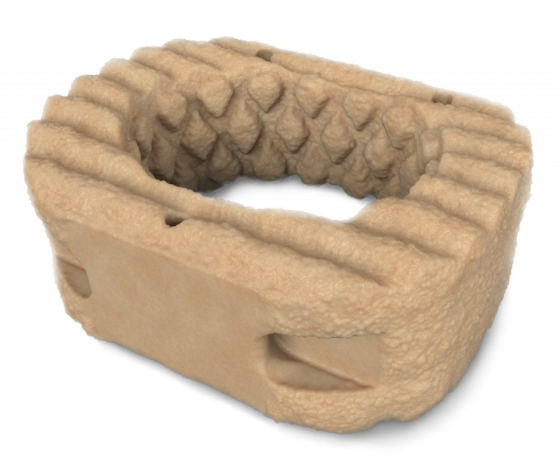
The Fortilink IBF devices are made using the company’s TETRAfuse 3D Technology which integrates 3D printed nano-rough surfaces for enhanced protein absorption, bioactivity, cell adhesion, and tissue growth for fusion in cervical and lumbar surgeries.
“While PEEK implants are widely used, data suggest PEEK has minimal characteristics to enhance implant osseointegration [the connection between living bone and a load-bearing artificial implant],” said Dr. Christopher Kepler, MBA, at the Rothman Orthopaedic Institute.
“The porous Fortilink cages with TETRAfuse 3D Technology have the potential to create a favorable environment for bone growth and are radiolucent. I look for the ongoing study to introduce surgeons to this product and demonstrate the advantages of Fortilink cages over other cage materials.”
The Clinical Evaluation of Fortilink Interbody Fusion Device with TETRAfuse 3D Technology, known as the FORTE study, is a post-market evaluation of the safety and performance of the Fortilink-C, -TS, and -L IBF implants. It ultimately seeks to access the radiographic evidence of cervical and lumbar fusion at three and six months post-surgery in DDD patients.
RTI Surgical defines DDD as an age-related condition when one or more discs between the vertebrae of the spinal column deteriorate or break down. Spinal fusion surgery is used as a common treatment option to reduce pain related to DDD.
Nevertheless, the company reports that health care costs and productivity losses associated with DDD-related surgeries are more than $50 billion annually. Thus, the FORTE study will assess the Forlink devices as an effective, cost-efficient solution. Dr. K. Brandon Strenge, at the Orthopaedic Institute,Shropshire, U.K., added:
“I’ve really been impressed with the TETRAfuse implants so far [and] I’m excited to see the bone growth showing on the CT scans, unlike the titanium implants out there.”
The data collected from this study will be collected from 150 patients from 20 U.S. medical facilities will be evaluated over the next 3.5 years. All of the DDD subjects are expected to be enrolled by Q1 2020.
Fortilink-C IBF System with TETRAfuse 3D Technology. Image via RTI Surgery.
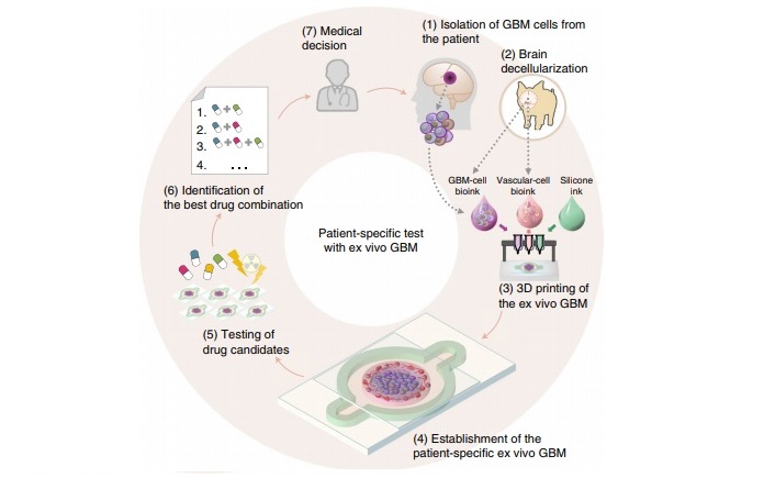
Glioblastoma, also known as glioblastoma multiforme (GBM), is an aggressive form of cancer found in the brain or spinal cord. In a study published in Nature Biomedical Engineering, the scientists detail how extracted GBM cells from cancer patients in an ex vivo model can emulate the characteristics of GBM human tumors. Professor Paek explained:
“[This] system confirms that cell-printing technology could be used to find a customized anticancer combination for each patient in the future treatment of brain cancer.”
3D bioprinting cancer cells
GBM is the most common form of brain cancer, accounting for approximately 50% of all malignant primary brain tumors, and is considered to be highly refractory (resistant) to chemoradiotherapy, according to the research.
A team of bioengineers, led by Professor Paek and Professor Cho sought to combat the ineffectiveness of such treatments by observing the isolated cancer cells from patients who had improved and deteriorating symptoms through chemotherapy.
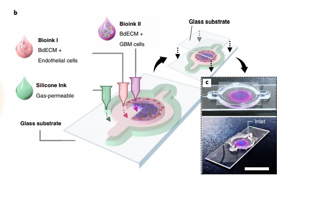
Bioink consisting of GBM and human vascular cells were printed sequentially to create a concentric ring structure on a chip which was also 3D printed from oxygen-permeable silicone. Using this chip the structural, biochemical and biophysical properties of the GBM tumors could be replicated.
Schematic illustration of the bioprinting and use of the patient-specific GBM-on-a-chip for the identification of an optimal drug combination for the patient. Image via SNUH/POSTECH.
The oral chemotherapy drug temozolomide was used to create a reaction in the cells similar to the clinical outcomes of chemotherapy. The research states that the “glioblastoma-on-a-chip reproduces clinically observed patient-specific resistances to treatment with chemoradiation.
“The model can be used to determine drug combinations associated with superior tumor killing for glioblastoma patients resistant to standard first-line treatment.”
“A bioprinted human-glioblastoma-on-a-chip for the identification of patient-specific responses to chemoradiotherapy” is co-authored by Hee-Gyeong Yi , Young Hun Jeong, Yona Kim, Yeong-Jin Choi, Hyo Eun Moon, Sung Hye Park, Kyung Shin Kang, Mihyeon Bae, Jinah Jang, Hyewon Youn, Sun Ha Paek , and Dong-Woo Cho.
Similarly, Shalini Guleria, Masters student at the University of Waikato, New Zealand, is 3D printing breast tumor cells to identify the best treatment for cancer patients. Last year, Guleria created a prototype tumor model using FFF 3D printing and is following this design in the development of an ex vivo bioprinted model.
B) Schematic illustration of the process for 3D printing the GBM-on-a-chip with various bioinks. C) Photographs of a mock GBM-on-a-chip. Image via SNUH/POSTECH.
Looking for a career in additive manufacturing? Visit 3D Printing Jobs for a selection of roles in the industry.
Featured image shows representative brain magnetic resonance images of the GBM patients. Image via SNUH/POSTECH.
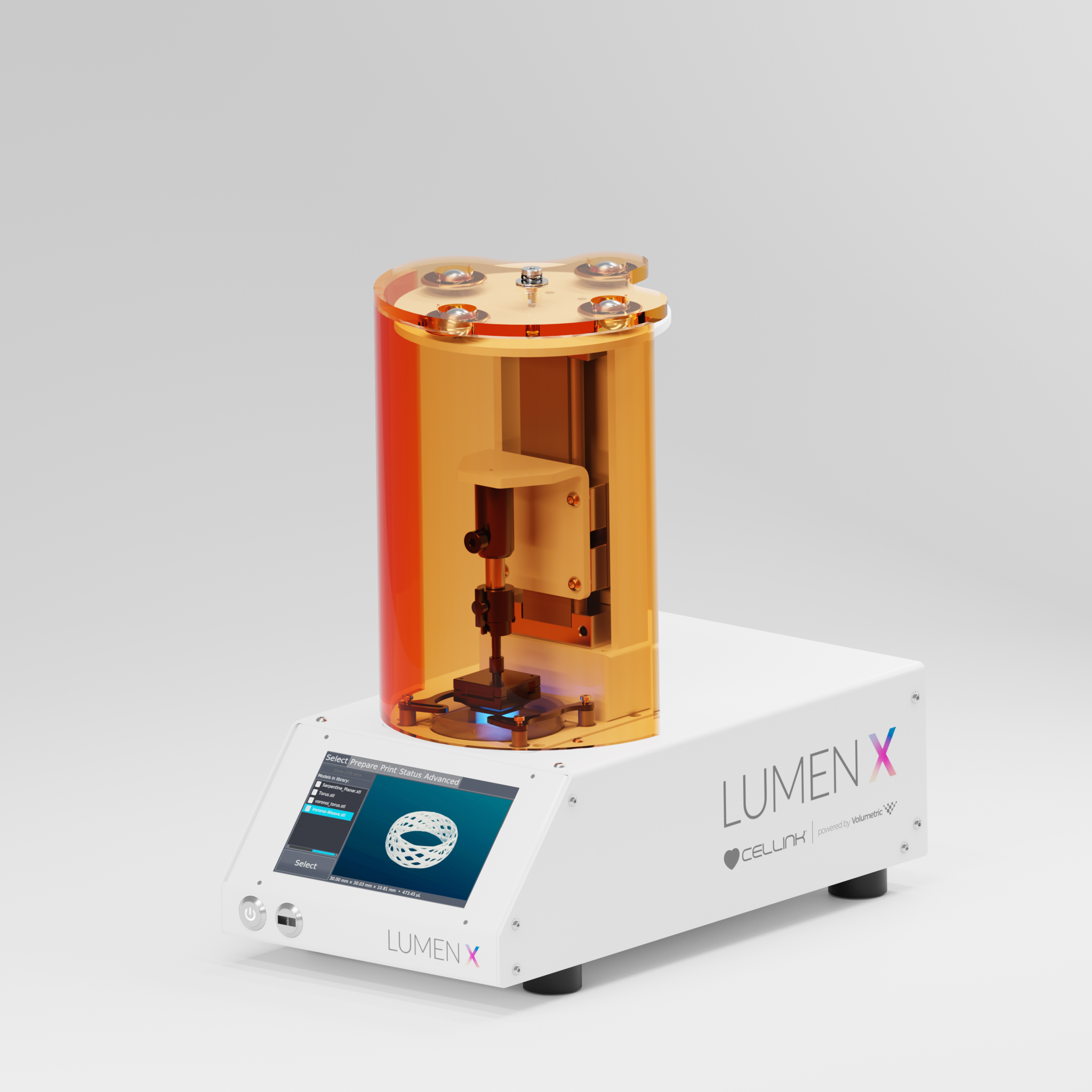
CELLINK, the Swedish manufacturer of the BIO X 3D printer, and Volumetrica Texas-based biomaterials and biomanufacturing company, have introduced the Lumen X Digital Light Processing (DLP) bioprinter to produce large vascular structures.
Designed as an entry-level platform, the Lumen X works with patent-pending bioinks to print high-resolution, macroporous, and vasculature structures, strengthening research regenerative medicine. Dr. Jordan Miller, the co-founder of Volumetric, said:
“Through this collaboration, we are harnessing the power and precision of light to structure living tissue through highly parallelized projection stereolithography and photopolymerization. We see applications in microfluidics, lab-on-a-chip devices, and vascularized living tissues containing millions of cells.”
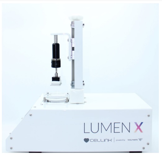
The Lumen X 3D bioprinter. Photo via CELLINK/Volumetric.
The Lumen X
Although Volumetric was only founded in 2018, co-founders Dr. Miller and Bagrat Grigoryan have spent five years researching and validating materials for 3D bioprinting “to empower the next generation of advanced biofabrication”.
Upon the launch of the Lumen X, Dr. Miller added, “We are so thrilled to see our research, the culmination of a combined 20 years of stereolithography and 3D bioprinting expertise, developed and commercialized through this incredibly strong partnership with CELLINK.”
The Lumen X has been established as a complementary product to the Holograph X and BIO X 3D bioprinters as well as all bioinks within CELLINK’s portfolio. It is said to leverage over one million points of light to bioprint microscopic features down to 200 microns.
This platform also photographically cures entire layers at once, crosslinking structures fifty times faster than alternative bioprinting methods. Moreover, the Lumen X is capable of print living cells within a BIO X-fabricated structure which and accelerate organ-on-a-chip research.
“We are excited to launch yet another product in our bioprinting technology portfolio,” said Erik Gatenholm, CEO and co-founder of CELLINK.
“This system will seamlessly complement our current product offerings of both hardware as well as bioinks and biomaterials. We look forward to continue offering the latest bioprinting technologies to our customers worldwide.”
The Lumen X 3D bioprinter. Photo via CELLINK/Volumetric.
Technical specifications and pricing
Printer Dimensions
24 x 43 x 41 cm (9.5 x 17 x 16.5 in)
Weight
9 kgs (20 lbs)
Technology
DLP
Pixel Resolution (XY)
50 µm
Projected Image
1280 x 800 pixels
Build Volume (maximum)
64 x 40 x 40 mm
Heated Platform
Maximum temperature of 37 °C
Z-Precision (motor-driven)
5 µm
Wavelength
405 nm Intensity
The price of the Lumen X has yet to be disclosed. However, a quote can be generated via the CELLINK website.
Looking for a change of pace or seeking new talent? Search and post3D Printing Jobs for opportunities and new talent across engineering, marketing, sales and more.
Featured image shows the Lumen X 3D bioprinter. Photo via CELLINK/Volumetric.
Gamma cameras are used to conduct functional scans of the brain, thyroid, lungs, liver, gallbladder, kidneys and skeleton. This medical imaging technique captures traces of radiation emitted by injected pharmaceuticals within cancer patients.
The 3D printed tungsten disks have been used to replace the lead components found in such devices. As detailed in the study published in European Journal of Nuclear Medical Imaging, the tungsten parts performed better for eliminating unwanted radiation, creating a clear final image.
“Gamma ray radiation is very powerful so you need heavy shielding to produce a clear image,” explained Dr. Jonathan Gear, Clinical Scientist in the Joint Department of Physics at the ICR and The Royal Marsden, and lead author of the study.
“Most gamma-ray cameras use thick lead, but denser materials like tungsten could be as effective while letting more useful radiation pass through to produce higher quality images.”
3D printing tungsten
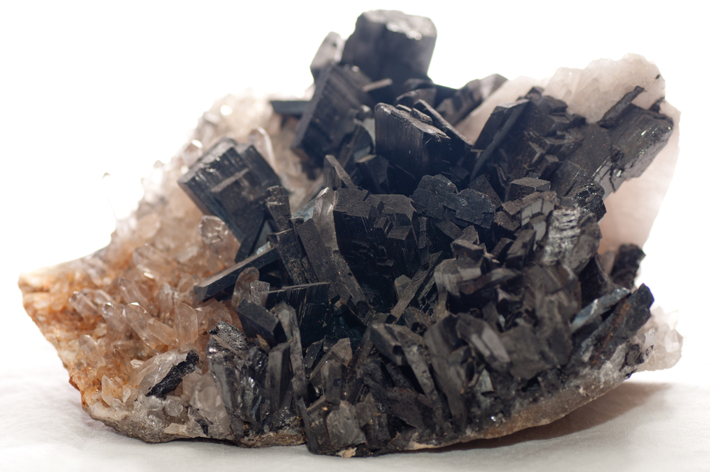
Found in the earthly mineral wolframite, tungsten is an inorganic, non-natural, rare metal compound. It is extremely dense and maintains the highest melting point of all metals, that is 3422 °C. With its beneficial material properties, including high tensile strength and high corrosion resistance, its use in additive manufacturing continues to be explored.
The 3D printed tungsten shields, known as collimators, integrated into gamma cameras by the ICR team were produced by Wolfmet a brand of M&I Materials, a Manchester-based specialist of industrial materials. Wolfmet created the shields using an EOS M280 3D printer using Selective Laser Melting (SLM).
A honeycomb structure of holes was used in these components to allow the adequate amount of radiation through to create an image while blocking unwanted radiation so the image is sharp. Dr. Gear added, “Our research shows that 3D printed tungsten could be used in gamma cameras, offering greater flexibility in the sorts of designs that could be used in the future.”
The element tungsten is found in the mineral wolframite. Photo via Shutterstock/farbled.
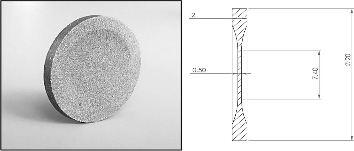
The 3D printed tungsten shields were 20% thicker than standard lead gamma camera collimators. The measured attenuation of a gamma beam using such shields was also seen to be lower than the value determined from the National Institute of Standards and Technology (NIST).
With a reduction in the strength of gamma radiation, the IRC researchers have concluded that tungsten collimators are feasible and superior to its lead alternative. Also with the use of additive manufacturing, the collimator can have a bespoke collimator design within gamma cameras.
“Characterisation of the attenuation properties of 3D-printed tungsten for use in gamma camera collimation” is co-authored by Jonathan I. Gear, Jan Taprogge, Owen White and Glenn D. Flux.
Photograph of the 3D printed disk and schematic diagram of disk dimensions. Image via ICR.
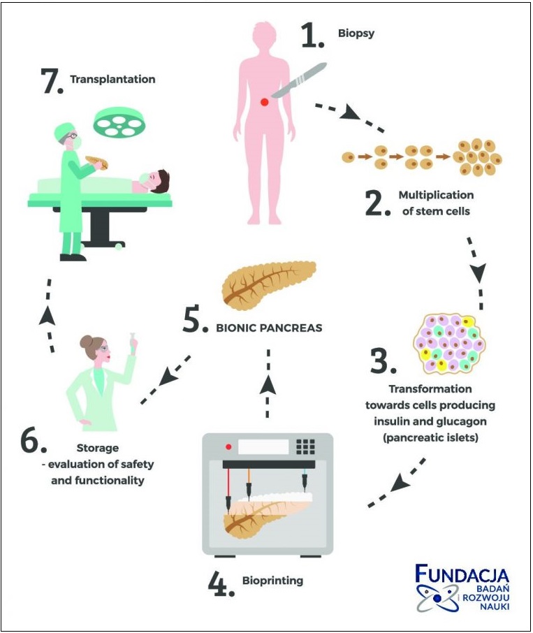
In an effort to combat diabetes, Michał Wszoła MD, Ph.D., Principal investigator at Fundacja BIRN, is leading a project to 3D bioprint scaffolds using pancreatic tissue or insulin-producing cells to form an artificial pancreas.
Although current methods of insulin production, that is, an insulin pump i.e., intensive insulinotherapy, and pancreas transplantation can treat type I diabetes, the scientists have identified limitations such as organ shortages and post-surgical complications.
“The goal of the project is to create a functional pancreas. One that can be transplanted to the patient without major problems,” said Professor Wszoła.
“It will enable people affected by diabetes to function normally, and more importantly prevent the development of secondary complications, which are the causes of most deaths.”
The process of the bionic pancreas. Image via Fundacja BIRN.
Creating a bionic pancreas
According to the World Health Organization (WHO) over 60 million people in European suffer from diabetes, and this number continues to increase. To address this problem, in 2015, Fundacja BIRN researchers established the BIONIC consortium, which includes embers such as Swedish 3D bioprinter and materials developer CELLINK.
“It is commonly known that the number of transplants is growing, the demand for organs exceeds the amount that can be obtained from donors,” added Professor Wszoła.
“In addition, after pancreas transplantation, the operation itself carries the risk of surgical complications, and the continuation of life after pancreas transplantation is associated with the continuous intake of immunosuppressive drugs.”
“A pancreas created from its own transformed stem cells eliminates these two major problems.”
3D bioprinting artificial pancreatic tissue
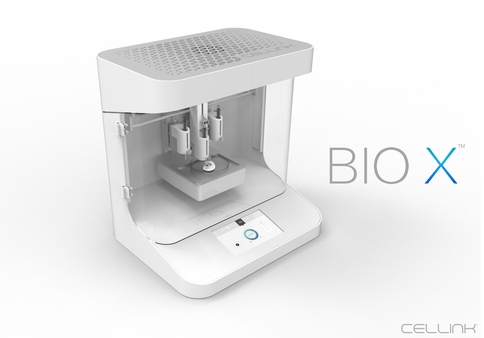
As of 2018, CELLINK’s Bio X bioprinter from CELLINK is being used at Fundacja BIRN to print living cells and pancreatic islet – a portion of tissue – which will eventually form a bionic pancreas. The project is now preparing for its first pre-clinical animal trials which will start later this year. Hector Martinez, co-founder and CTO, explained, “We are so extremely excited about this, especially since the application is printing a pancreas.”
“There is such major need for artificial pancreatic tissue today and the development has not been moving fast enough. While we do understand that this will take some time, we do recognize that this is a tremendously important application.”
Also, visit 3D Printing Jobs for new opportunities in your area.
Featured image shows a pancreas puzzle concept: hands of a surgeon with surgical instruments (tools) perform pancreas surgery as a result of acute or chronic pancreatitis, pancreatic diabetes, pancreatic cancer. Image via Fundacja BIRN.