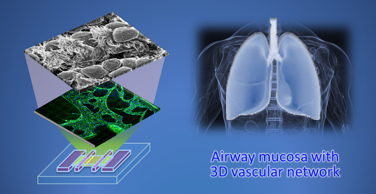
Korean researchers from Pohang University of Science and Technology (POSTECH) and Seoul National University (SNU) have used 3D bioprinting to create a biomimetic airway-on-a-chip.
This device, equipped with a naturally-derived blood vessel network, emulates biochemical responses and is used to study respiratory diseases. In the research article published in Biofabrication, Ju Young Park, lead author of the study explained:
“We reproduced an in-vivo-like 3D vascular network by assembling endothelial cells and fibroblasts using the dECM [decellularized extracellular matrix] bioink in a one-step printing process.”
“The structure we produced has the same physiological functions as the biological airway epithelium [surface tissue] and so can be used to model diseases like asthma.”
3D bioprinting respiratory systems
The research team was inspired to develop life-like airway models after observing the rising rate of air pollution, globally, which contributes to the number of people suffering from respiratory diseases.
With such airway-on-a-chip models, the researchers can observe and characterize inflammatory diseases, including asthma, chronic lung disease, and rhinosinusitis – an inflammation of the sinuses and nasal cavity.
According to Park, the human airway is lined with surface cells known as epithelial. These cells mainly contain ciliated goblet and basal cells which connect with a basement membrane. In addition, under this membrane is Lamina propria, a thin layer of loose connective tissue, which contains blood vessels and stromal fibroblasts cells.
“To mimic this complex 2D/3D structure and the cellular composition of the airway mucosa, we assembled a 2D airway epithelium on a 3D vascular platform,” added Park.
“We reconstructed the natural 3D vascular network by 3D cell printing the dECM bioink containing endothelial cells and fibroblasts.”
Biomimetic 3D cell structures
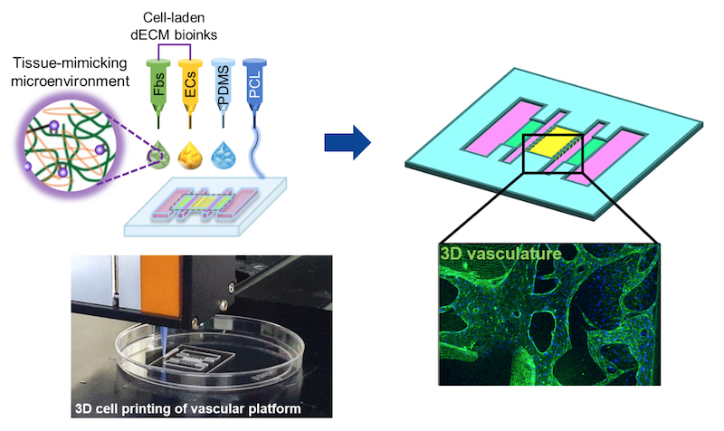
Using an in-house 3D bioprinter equipped with six dispensing heads, the team were able to successfully produce complex microstructures replicating the fine cellular arrangement in natural tissue.
“The other four printing heads operate on a three-axis motorized stage and we control their movement using computer programs,” stated Park. “Two of the printing heads were connected to a pneumatic pressure-based system that dispenses a synthetic polymer to fabricate the supporting framework for the airway.”
Following their research, the team concluded, “that pathological interaction between the airway endothelium and the vascular network in the airway are reproducible in [the] airway-on-a-chip model, and that the exacerbation of inflammatory responses by the vascular network in vivo is also reproducible in vitro.”
As such, the team believes the 3D cell printed airway-on-a-chip will be useful for animal models in analyzing diseases and testing the efficiency of drugs in the preclinical phase.
This technology will be commercialized by T&R Biofab, a Korean biotechnology company.
The research article, “Development of a functional airway-on-a-chip by 3D cell printing,” is co-authored by Ju Young Park, Hyunryul Ryu, Byungjun Lee, Dong-Heon Ha, Minjun Ahn, Suryong Kim, Jae Yun Kim, Noo Li Jeon, and Dong-Woo Cho.
Submit your nominations now for the 3D Printing Industry Awards 2019.
Also, for the latest medical 3D Printing Industry news subscribe to our newsletter, follow us on Twitter and like us on Facebook.
Looking for a fresh start in the new year? Visit 3D Printing Jobs to commence your new career in additive manufacturing.
Featured image shows a depiction of the 3D printed airway-on-chip. Image via Ju Young Park/ Seoul National University.
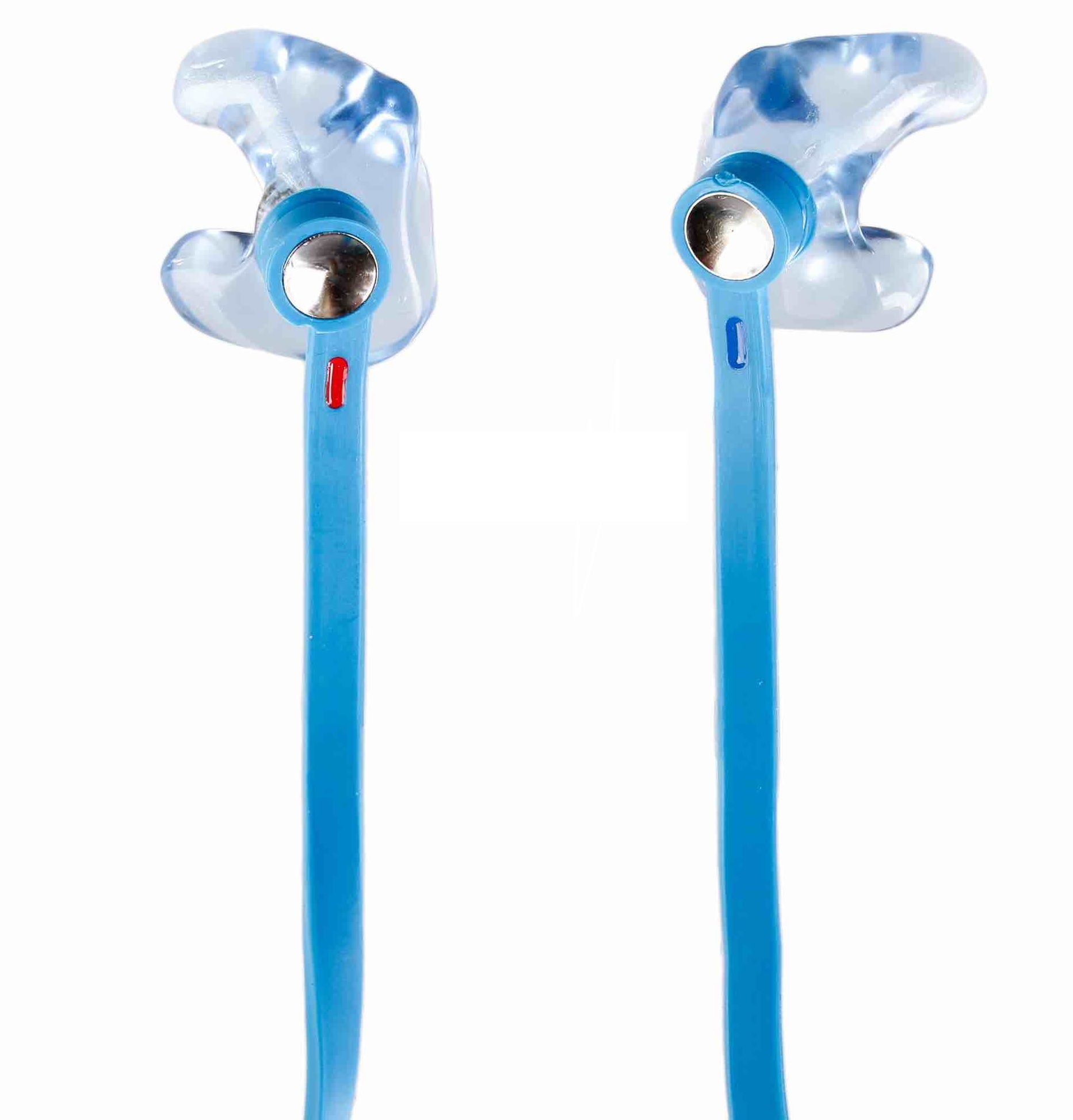
Prodways Group, a French 3D printer producer and service provider, has announced the acquisition of hearing aid manufacturers Surdifuse and L’Embout Français.
Upon acquiring 100% of the companies share capital, Prodways Group plans to accelerate production of customized 3D printed medical devices such as customized ear tips for hearing aids.
Accelerating 3D printed hearing aid production
In 2017, the two historic French laboratories, Surdifuse and L’Embout Français, merged to form Surdifuse-L’Embout Français. Guillaume Felten, President of Surdifuse-L’Embout, explained, “The hearing market is undergoing significant change. We are structured to continue to serve our customers in the best conditions of quality, time and order tracking.”
“Cross-cutting exchanges of good practices, methods, management tools and the complementarity of production sites will allow us to gain considerable time to achieve this goal. This is all the meaning of this rapprochement and I have a lot of ambitions for this Group.”
By 2018, Surdifuse-L’Embout Français became the French leader in the tailor-made adapter market for hearing aids. In the same year, it produced approximately 150,000 hearing aids using additive manufacturing and 3D modeling.
Prodways 3D printed hearing aids
Preceding this acquisition, Prodways Group previously acquired a 75% stake in medical device manufacturer Interson-Protac that also uses 3D printing to make hearing aids. Prodways Group will now strategically lead Surdifuse-L’Embout Français to strengthen its place in the medical sector, which the company has identified as a major growth driver in the Products division.
In addition, Prodways Group has accrued 40 new employees who will work over sites in Paris and Lyon. This acquisition is expected to generate revenue of more than €3 million and make a positive contribution to the Group’s income.
Submit your nominations now for the 3D Printing Industry Awards 2019.
Also, for the latest medical 3D Printing Industry news subscribe to our newsletter, follow us on Twitter and like us on Facebook.
Looking for a fresh start in the new year? Visit 3D Printing Jobs to commence your new career.
Featured image shows 3D printed hearing aids from Surdifuse-L’Embout Français. Photo via Surdifuse-L’Embout Français.
Anatomiz3D Medtech Private Limited, a Mumbai-based medical 3D printed service bureau has partnered with fellow Indian bureau, 3D Incredible, to provide patient-specific surgical solutions.
Following two years of collaborations in the medical and dental sector, 3D Incredible has now invested an undisclosed amount sum into Anatomiz3D to form a one-stop shop for customized healthcare.
The companies have previously worked on multiple cases of cranial, maxillofacial and orthopedic 3D printed implants.
India integrates 3D printing in healthcare
Founded in 2017, 3D Incredible stemmed from India’s iron powder manufacturer Industrial Metal Powders (IMP), a company established in 1974. The company is utilizing this experience to implement metal 3D printing technology to cater to the engineering and medical industries.
IMP remains the only company in India to receive ISO 9001 and ISO 13485 certifications for the production of medical devices. This has led to the development of multiple custom 3D printed implants in neuro, maxillofacial and orthopedic surgeries across the country.
Anatomiz3D, on the other hand, began as the healthcare vertical of Sahas Softech, a Mumbai-based design studio. The company now provides 3D modelling from 2D patient CT and MRI data as well as additive manufacturing to create 3D printed surgical guides.
Three years ago, Anatomiz3D reportedly became the first in India to provide surgeons with a patient-specific Paediatric Cardiology model prior to surgery; the company has now expanded into similar fields as its partner including Neurosurgery, Oral and Maxillofacial, Head and Neck, Spine, and Orthopaedics.
Together, the companies aim to bring additive manufacturing technologies to the masses while developing cost-effective, precise, and high-quality 3D printed medical products.
Additive manufacturing and the healthcare sector
3D printed surgical guides have aided numerous patient case studies around the world. In November, 3D Systems began its partnership with non-profit organization OpHeart to produce 3D printed anatomical models used in delicate surgeries involving babies with life-threatening heart defects.
Prior to this, researchers at the Great Ormond Street Hospital (GOSH) in London began developing 3D printed replicas of children’s hearts for surgeons to better plan complex and crucial heart surgeries.
Moreover, 3D printed medical implants and healthcare services are finding increasing use by healthcare professionals. As a result, the Belgian Health Care Knowledge Center (KCE) published a report of recommendations for the “Responsible use of High-Risk Medical Devices” made using 3D printing.
Submit your nominations now for the 3D Printing Industry Awards 2019.
Also, for the latest medical 3D Printing Industry news, subscribe to our newsletter, follow us on Twitter and like us on Facebook.
Looking for a fresh start in the new year? Visit 3D Printing Jobs to get a head start.
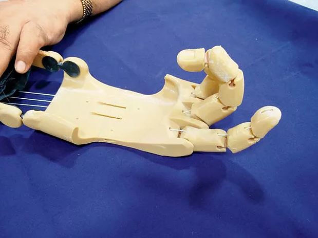
Featured image shows a 3D printed hand prosthetic created by Anatomiz3D. Photo via Anatomiz3D.
The month of August welcomed various medical innovations within the 3D printing industry. The automotive and aerospace sector also further integrated additive manufacturing through Bugatti’s latest supercar, the Divo, and NASA’s cube satellite set for space from the Hindustan Institute of Technology and Sciences.
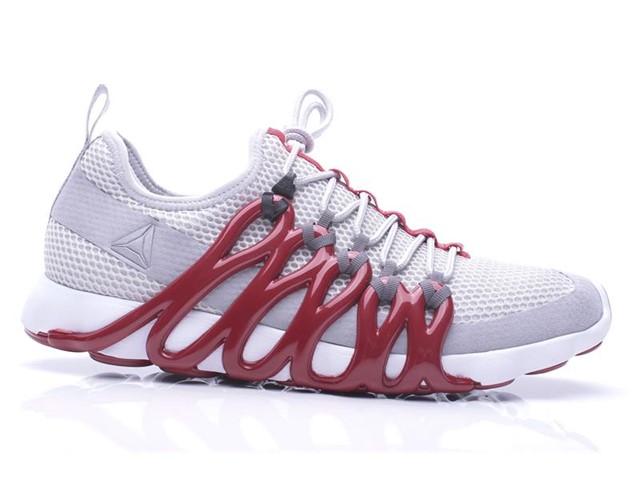
Moreover, Reebok and BASF began production of its 3D printed sneaker, the Liquid speed.
Medical 3D printing innovations
In a proof-of-concept project, the United States Department of Veterans Affairs (VA) for military healthcare began applying 3D printing to develop artificial lungs. Upon developing 3D printed chips devices for organs, Dr. Joseph Potkay, a biomedical engineer at the VA Ann Arbor Health Care System, Michigan, said:
“The flexibility in design afforded by 3D printing gives us more freedom and thus the ease to build artificial lungs with a small size and pressure drops that are compatible for operation with the body’s natural pressures.”
In other news, the University of Sydney’s Save Sight Institute received a $1.15 million (AUD) grant from the New South Wales (NSW) Government’s Medical Devices Fund (MDF) to progress commercialization of its 3D corneal biopen.
Dubbed as the “iFix Pen”, the hand-held co-axial 3D printer is capable of extruding bioink directly onto an eye to aid in the regeneration of cells on corneal ulcers. The pen also creates a biological barrier towards ongoing cornea damage caused by infections.
Similarly, the McAlpine Research Group from the University of Minnesota (UMN) successfully 3D printed optoelectronic devices using polymer photodetectors on hemispherical surfaces. This development was marked as a significant step towards “bionic eyes” to aid the visually impaired.
Investments and aerospace
Within business, the city of Querétaro, Mexico, became the host of the first Additive Manufacturing Consortium (Conmad) in Latin America. Investing over $13 million, the Conmad includes Cinvestav, Mexico’s National Center for Research and Advanced Studies, CIATEQ an Advanced Technology Center, and CIDESI, a Center for Engineering and Industrial Development.
The main aim of Conmad is to validate and explore viable 3D printing technology for production to implement new additive manufacturing infrastructure in North-Central Mexico.
Additionally, Digital Alloys, a Boston based developer of metal 3D printers, received a $12.9 million investment during a series B financing including G20 Ventures, Boeing HorizonX Ventures, Lincoln Electric, and Khosla Ventures. This financing is being used to develop Digital Alloys’ new metal additive manufacturing approach called Joule Printing.
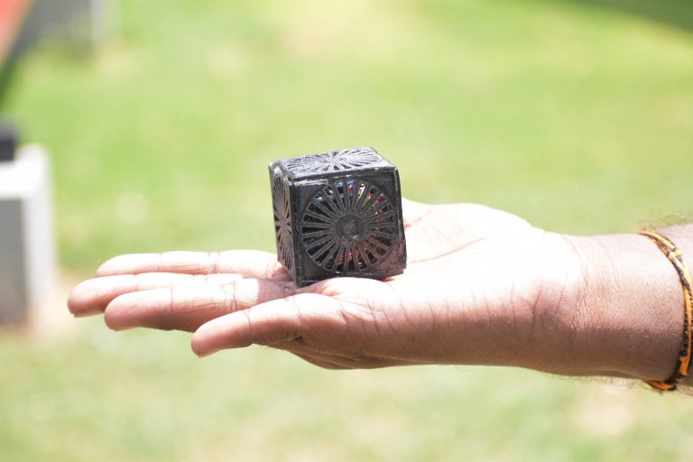
Developing one of the lightest satellites in the world, Indian 3D printer manufacturer 3Ding, and students at the Hindustan Institute of Technology and Sciences, created a 3D printed a cube satellite set for launch on a NASA Columbia Scientific Balloon Facility at Fort Sumner, New Mexico.
Known as Jai Hind 1-S, the cube, weighing 33.3 grams, was crafted as a submission for Cubes in Space, a free, global competition for students ages 11-18.
The Liquid Factory
Causing excitement in both the 3D printing and fashion industry, Reebok and its materials provider BASF released details about its Liquid Factory sneaker 3D printing technology. Acting as a step towards production, Reebok opened a Rhode Island facility where 3D printed shoes will be made.
The Bugatti Divo
Also drawing attention to the capabilities of additive manufacturing at the Quail, A Motorsports Gathering.was the Bugatti Divo. With a price tag of €5 million, the Bugatti Divo supercar displayed 3D printed fin taillights, which were designed for optimum handling performance on winding roads.
“The Divo is a further example of our design philosophy ‘Form follows Performance‘. In this case, the engineers and designers aimed to create a vehicle focusing on cornering speeds and lateral dynamics,” said Achim Anscheidt, Director of Design at Bugatti.

Facebook’s stance on 3D printed guns
Contributing to the debate over 3D printed guns, social media giant Facebook became the latest organization to weigh in on the discussion in an update to its Community Guidelines. The platform stated: “Sharing instructions on how to print firearms using 3D printers is not allowed under our Community Standards,” much to Cody Wilson’s dismay.
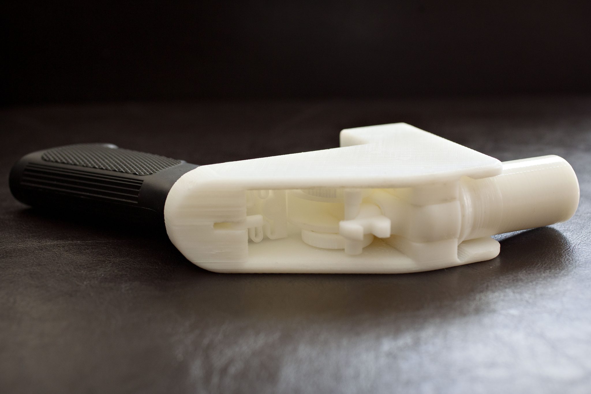
Finally, in hardware developments, Aleph Objects, the Colorado-based manufacturer of the LulzBot range of desktop 3D Printers, unveiled a new, high-precision tool head at SIGGRAPH 2018 in Vancouver – the LulzBot Aerostruder v2 Micro.
Who made the best contribution to the 3D printing industry? Make your nominations now for the 3D Printing Industry Awards 2019.
Also, for the latest 3D Printing Industry news throughout 2019 subscribe to our newsletter, follow us on Twitter and like us on Facebook.
Looking for a fresh start in the new year? Visit 3D Printing Jobs to get a head start.
Featured image shows the Bugatti Divo, complete with 3D printed taillights.
Researchers from Pennsylvania State University have created a novel 3D printing method to create tissue building blocks with micropores.
According to the Penn State team, micropores in artificial tissues forming bone and cartilage allow nutrient and oxygen diffusion into the core. By 3D printing stem cells derived from human fat mixed with sodium alginate porogens found in seaweed, the materials can be extruded into tiny particles that, when dissolved, leave behind pores in the fabric of the tissue.
Such additively manufactured porous structures allow both nutrients and other fluids to circulate and demonstrate the potential for lab-grown tissue containing blood vessels. Ibrahim T. Ozbolat, associate professor of engineering science and mechanics, at Penn State said:
“Cells die if nutrients and oxygen can’t get inside. One of the problems with fabrication of tissues is that we can’t make them large in size.”
“These patches can be implanted in bone or cartilage. They can be used for osteoarthritis, patches for plastic surgery such as the cartilage in the nasal septum, knee restoration and other bone or cartilage defects.”
3D printing tissue with micropores
Considered as an alternative to vascularization, the researchers are creating tissue building blocks with micropores in an effort to grow blood vessels within tissue. The outcome has been dubbed as porous tissue strands.
Using the stem cells mixture, the researchers 3D printed strands of undifferentiated tissue which then combined to form patches of tissue. The tissue was then exposed to a chemical solution which converts the stem cells into specific cells, in this case, bone or cartilage.
As cartilage does not contain blood vessels, porous structures can be easily created to produce implants with natural porosity. Currently, only tiny patches can be made, however, in a recent issue of Biofabrication, the researchers reported that the strands maintained 25% porosity and have pore connectivity of 85% for at least three weeks.
The researchers at Penn State involved in this study include Yang Wu, Monika Hospodiuk, Hemanth Gudapati, Thomas Neuberger, Srinivas Koduru, and Dino J. Ravnic, as well as Weijie Peng, from the department of pharmacology at Nanchang University, China.
Submit your nominations now for the 3D Printing Industry Awards 2019.
Also, for the latest medical 3D Printing Industry news throughout 2019 subscribe to our newsletter, follow us on Twitter and like us on Facebook.
Looking for a fresh start in the new year? Visit 3D Printing Jobs to get a head start.
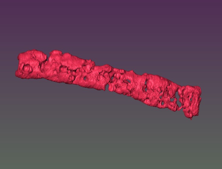
Featured image shows a reconstructed 3D image of porous tissue strand using magnetic resonance imaging. Image via Ozbolat laboratory/Penn State.
Scientists from the Indian Institute of Technology (IIT) Delhi and IIT Kanpur have created a 3D bioprinting method to develop load-bearing bone cartilage.
In a study published in the scientific journal, ACS Biomaterials Science & Engineering, the researchers took inspiration from the biological process within the human body where stem cells form into bones (osteoblasts), to create a 3D printed bone construct.
“A major challenge in bone tissue engineering is to develop clinically conformant load-bearing bone constructs in a patient-specific manner,” the paper states.
“3D bioprinted silk-gelatin constructs enabled adequate cellular attachment, proliferation and most importantly, articular cartilage differentiation for activating pathways of organogenesis [organ production] in a patient-specific manner.”
Forming 3D printed bone constructs
According to the study, load-bearing, long bones, such as the femur, are produced from a process involving stem cells which form cartilage templates. These templates experience further differentiation to form bone cells which are designed to bear weight.
According to Sourabh Ghosh a Professor from the Department of Textile Technology at IIT Delhi, attempts to develop load-bearing bones using different scaffolds have bypassed the template stage of cartilage formation to differentiate stem cells directly into bone cells.
“The efficacy of such bone constructs is yet to be demonstrated in bearing loads. There is a very poor correlation between bone constructs developed in vitro and in vivo. Also, the gene expression pattern of these tissue-engineered bones largely differs from human adult bone.”
Using bioink containing silk proteins, mesenchymal stem cells, a thyroid hormone (Triiodothyronine), and growth factors, as well as a 3D bioprinter, the research team were able to engineer bone cartilage.
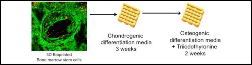
Load-bearing stem cells
Professor Ghosh explained, “We followed a four-step process to develop the load-bearing bone. We first developed chondrocytes (cartilage) from stem cells and then differentiated them into hypertrophic chondrocytes [specialized cells for bone growth].”
“During this process, the sponge-like cartilage becomes a brittle tissue. Here it is following the development biology mechanism to become a bone.”
The hypertrophic chondrocytes then differentiate into osteoblasts prior to them becoming adult bone cells known as osteocytes. Professor Amitabha Bandyopadhyay from the Department of Biological Sciences and Bioengineering at IIT Kanpur further explained:
“Compared to bone formed directly from stem cells, the extracellular matrix of the bone construct developed through the intermediate cartilage process was 10s of times higher.”
“The load-bearing capacity of a bone depends primarily on the quality of extracellular matrix. In loading-bearing bones, the extracellular matrix comprises 95% while bone cells are just 5%. So if you are trying to fabricate a load-bearing bone construct it is better to have more extracellular matrix.”
The researchers have deduced that the bone construct demonstrated better results than the bone developed directly from stem cells. The team intends to undertake studies on animals.
The research paper, “Developmental Biology-Inspired Strategies To Engineer 3D Bioprinted Bone Construct” is co-authored by Shikha Chawla, Aarushi Sharma, Amitabha Bandyopadhyay, and Sourabh Ghosh.

Submit your nominations for the 2019 3D Printing Industry Awards here.
Also, for all the latest 3D printing news, subscribe to the 3D Printing Industry newsletter, follow us on Twitter, and like us on Facebook.
Make your next additive manufacturing career move or hire new talent. Search and post 3D Printing Jobs on our free jobs service.
Featured image shows an X-ray of cartilage damage in the knee. Image via medicalnewstoday,
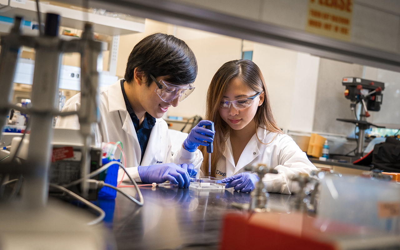
Bioengineers from the University of California San Diego (UCSD) have developed a 3D bioprinting method that integrates natural materials which produce lifelike organ tissue models.
The UCSD team used their method to create blood vessel networks capable of keeping a breast cancer tumor alive outside the body as well as a model of a vascularized human gut. The research, recently published in Advanced Healthcare Materials, aims to accelerate the production of human organ models to be studied for pharmaceutical drug screening.
“We want to make it easier for every day scientists—who may not have the specialization required for other 3D printing techniques—to make 3D models of whatever human tissues they’re studying,” said first author Michael Hu, a bioengineering Ph.D. student at the UC San Diego Jacobs School of Engineering.
3D printing a living blood vessel network
Last year, UCSD researchers additively manufactured a framework of functional blood vessels using a Digital Light Processing (DLP) method with hydrogel and encapsulated cells. Recognizing the importance of blood vessel networks in transporting blood, nutrients, and waste around the human body, UCSD engineers also developed 3D printed tissue that mimics the liver in terms of structure and function.
Now, UCSD scientists have developed created an “easy-to-use” technique to produce long‐term culturable ex vivo vascularized tissues. Using a commercial 3D printer, the researchers are able to print a scaffold out of a water-soluble material known as polyvinyl alcohol. Following this, a thick coating made of natural materials is poured over the scaffold which is then cured, solidified, and then flushed out the scaffold material inside to create hollow blood vessel channels.
The insides of the channels are then coated with endothelial cells (cells that line the insides of blood vessels). Cell culture media is then inserted through the vessels to keep the cells alive and growing.
“The models would be more advanced than standard 2D or 3D cell cultures, and more relevant to humans when it comes to testing new drugs, which is currently done on animal models,” added Hu.
Stimulating artificial organs
The 3D bioprinted blood vessels are made from materials such as fibrinogen – a glycoprotein that circulates in blood – as well as matrigel, a gelatinous protein mixture required for optimal growth of cell culture.
“We wanted to use materials that were natural rather than synthetic, so we could make something as close to what’s in the body as possible. They also needed to be able to work with our 3D printing method,” explained Bioengineering undergraduate student Xin Yi (Linda) Lei, a co-author on the study.
Following successful experimentation of the printed blood vessels within a breast cancer tumor tissues, Hu concluded:
“Our hope is that we can apply our system to make tumor models that can be used to test anti-cancer drugs outside the body. Breast cancer is one of the most common cancers—it has one of the largest portions of research dedicated to it and one of the largest panels of pharmaceuticals being developed for it. So any models we can make would be useful to more people.”
“Facile Engineering of Long-Term Culturable Ex Vivo Vascularized Tissues Using Biologically Derived Matrices,” is co-authored by Michael Hu, Amir Dailamy, Xin Yi Lei, Udit Parekh, Daniella McDonald, Aditya Kumar, and Prashant Mali.

Stay updated with the latest medical innovations in additive manufacturing by subscribing to our free newsletter. Also, follow us on Twitter and like us on Facebook.
Join 3D Printing Jobs now to search for the next step in your career.
Featured image shows bioengineering graduate student Michael Hu and undergraduate student Xin Yi (Linda) Lei constructing a vascularized gut model using their team’s new 3D bioprinting technique. Photo via David Baillot/UC San Diego Jacobs School of Engineering.
Clinicians of various specialties require autologous implantable tissues to repair and replace damaged tissue caused by diseases or trauma. With 3D bioprinting it is possible to manufacture such tissues, however, its biomechanical characteristics reduce its capabilities to be sutured.
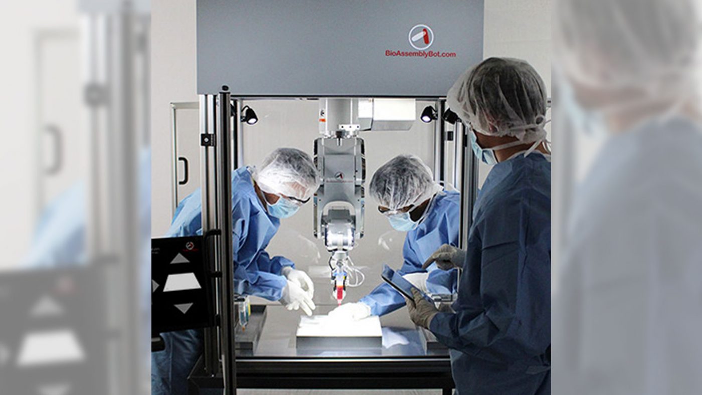
As a result, 3D.FAB, a French additive manufacturing platform, is developing a “living bandage” using 3D bioprinting and direct additive manufacturing. According to the platform’s project “STRESSKIN,” such technologies can be positioned in this field of regenerative and personalized medicine to address this issue.
The STRESSKIN Project
3D.FAB focuses on technological academic innovation in the field of health. In particular, the platform holds two main areas of expertise; the first includes biochemistry, especially diagnosis with prototyping 3D lab-on-chip, novel materials for 3D medical devices, biocompatible polymers, and cell-size 3D printing. The second attends to regenerative medicine through bioprinting living tissues.
One of several private projects from 3D.FAB, STRESSKIN aims to create innovative solutions to elevate the mechanical properties of bio-printed tissues towards biomimetic properties. “Once acquired, the applications will be vast and allow a multitude of specialists to find autologous and sustainable solutions to replace and repair tissue,” states 3D.FAB.
The 3D printed bandage is from a cell-based bioink. Using a BioAssemblyBot, a 6-axis robotic arm for biofabrication, this material is deposited onto the patient’s skin, forming an autograft that will create new skin in approximately two weeks. This approach is said to overcome other 3D printed skin solutions which have been proven to be too fragile to be sutured.
3D printed bandages
3D printed medical bandages are finding increasing uses by healthcare professionals. Scientists have developed smart bandages to treat and monitor open wounds and burns, ultimately accelerating the healing process.
Last year, Swansea University’s Institute of Life Science integrated 3D printing, nanotechnology, and biochemistry to create a fabric laced with minute particles of electro-conductive ink, for such injuries. This particular smart bandage collects data which is used to determine whether a wound needs extra care.
Following this, researchers at MIT debuted its perforated elastomer film with “kirigami” slits using 3D printed molds to improve the adhesiveness of additively manufactured medical bandages and wearable electronics.
Keep up with the latest additive manufacturing medical developments by subscribing to our free newsletter. Also, follow us on Twitter and like us on Facebook.
Join 3D Printing Jobs now to search for the next step in your career.
Featured image shows the development of the living bandage. Clip via 3D.FAB.
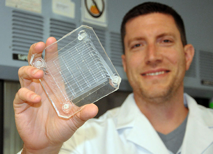
NewPro 3D, a Vancouver-based Direct Light Processing (DLP) 3D printing technology company, has partnered with Materialise, a Belgian software provider, to accelerate the additive manufacturing process for medical models.
With NewPro 3D’s NP1 3D printer, the companies plan to offer a one-stop solution for file preparation that can convert MRI and CT scan data to an STL file for additive manufacturing in approximately 10-15 minutes.
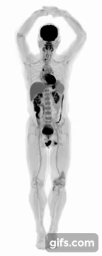
Elsewhere, researchers at the University of California (UC) Davis have designed the first medical 3D imaging full body scanner capable of producing an image in approximately a single second.
NewPro 3D and Materialise
Earlier this year, NewPro 3D introduced the NP1 3D printer. This system uses the company’s patented Intelligent Liquid Interface (ILI) Technology to print end-use materials at a fast pace. ILI is a transparent wettable membrane between the photo-curing resin and the light source. NewPro 3D has designed the membrane to chemically enable faster movement between cured layers.
This technology eliminates the mechanical processes used on conventional additive manufacturing techniques allowing objects to be produced at “record-breaking speeds.” At RAPID + TCT 2018, the NP1 was shown to 3D print a 22cm long midsole in 2 hours and 10 minutes, a fraction of the time taken using SLA.
Following its collaboration with Materialise, NewPro 3D’s Beta phase, the improved NP1 printers have been tested by the Radiology Department at Stanford University, the University of Louisville School of Dentistry, and Division of Cardiology at the University of Washington. Gabriel Castanon, COO at NewPro 3D said:
“We are now fully confident in the NP1 system [and] we will be delivering to customers in 2019. It is an exciting time for 3D printing in medical.”
First total-body 3D scanner
UC Davis scientists Simon Cherry and Ramsey Badawi have developed EXPLORER, a combined positron emission tomography (PET) and X-ray computed tomography (CT) scanner that can create 3D images of the entire body simultaneously.
The machine is made to capture radiation more efficiently than other scanners and can produce movies that can track specially tagged drugs as they move around the entire body. The scanner was developed in partnership with United Imaging Healthcare (UIH), based in Shanghai, who aims to eventually manufacture the devices for the broader healthcare market.
The first images from the scanner were shown at the Radiological Society of North America meeting last week. Badawi, Chief of Nuclear Medicine at UC Davis Health and Vice-Chair for research in the Department of Radiology explained:
“The level of detail was astonishing, especially once we got the reconstruction method a bit more optimized. We could see features that you just don’t see on regular PET scans. And the dynamic sequence showing the radiotracer moving around the body in three dimensions over time was, frankly, mind-blowing. There is no other device that can obtain data like this in humans, so this is truly novel.”
The researchers are currently using the EXPLORER to in medical studies to demonstrate how it can benefit patients and contribute to inform others of the whole human body in health and disease.
Stay updated with news related to 3D printing and the healthcare sector by subscribing to our free newsletter. Also, follow us on Twitter and like us on Facebook.
Join 3D Printing Jobs now to search for the next step in your career.
Featured image shows DLP technology from NewPro 3D. Photo via NewPro 3D.
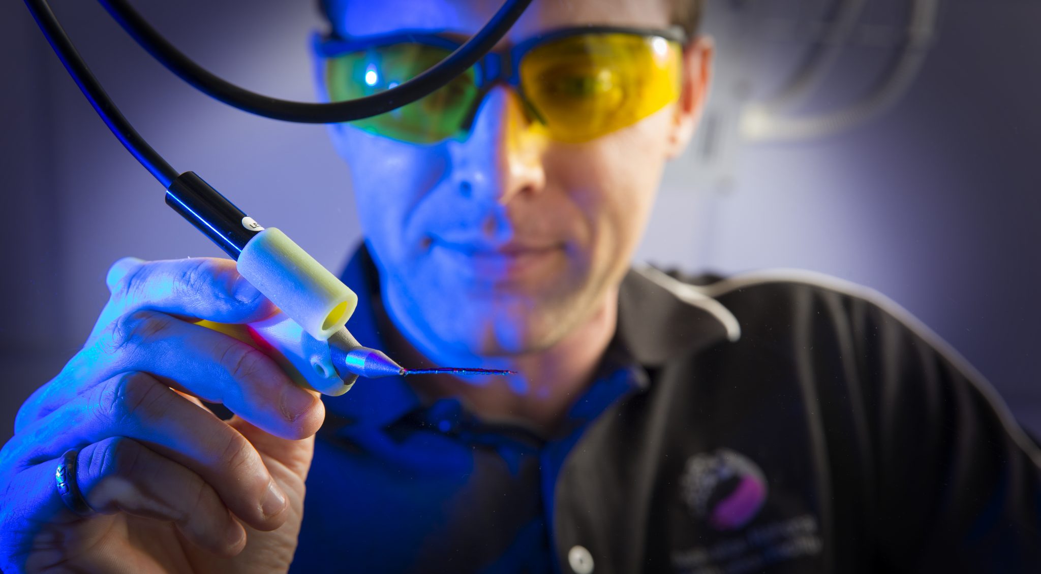
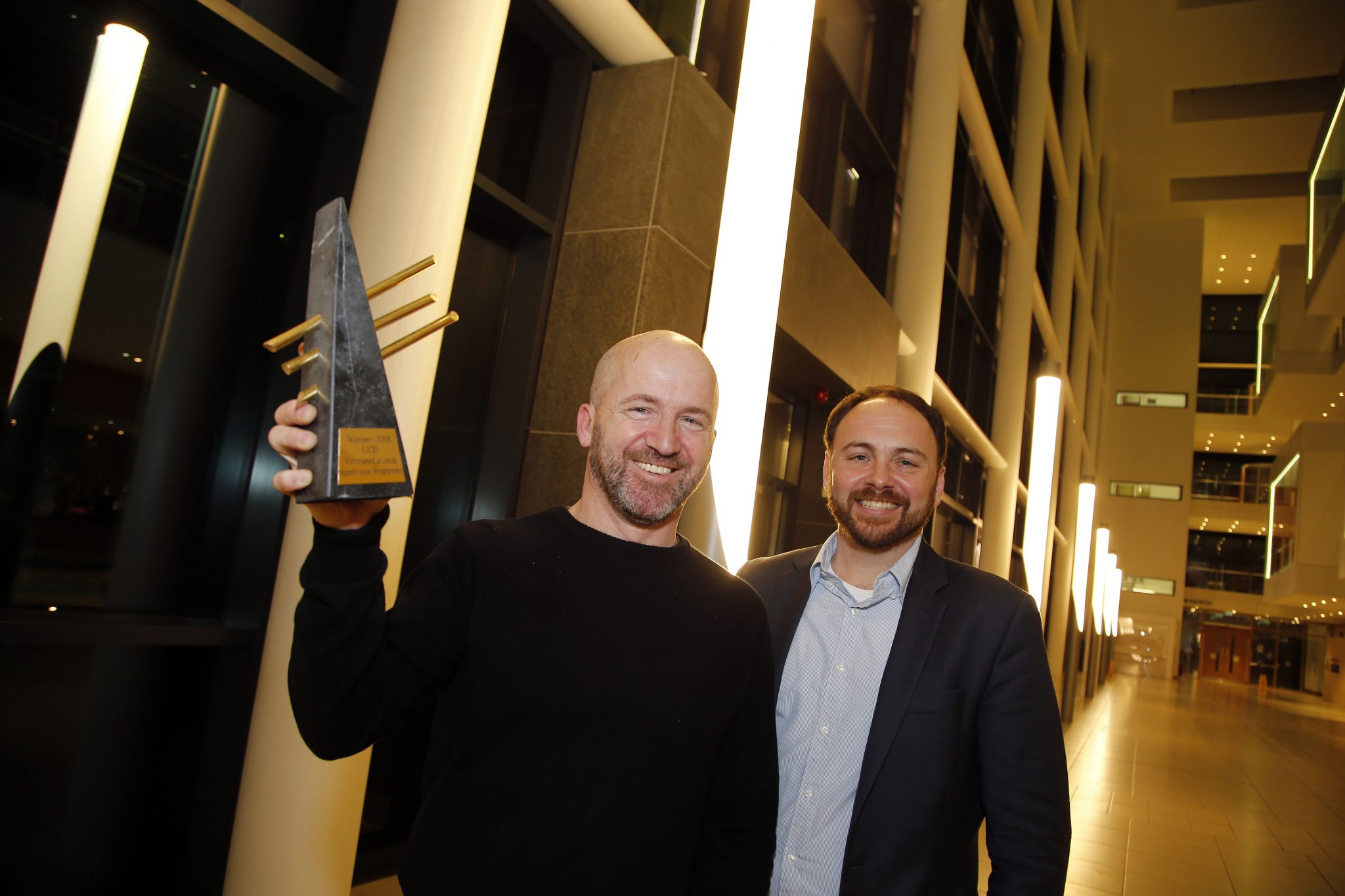
Naiad, an Irish 3D biotechnology company, has won University College Dublin’s (UCD) 2018 Start-Up of the Year Award. The company also received a €32,000 prize fund, as part of the 2018 UCD Venture Launch Accelerator Programme.
As one of six emerging UCD start-ups, Naiad is developing a novel liquid-based 3D bioprinter that helps researchers fabricate highly-reproducible and realistic 3D tissue models.
“We are strongly committed to delivering impact from our research and innovation activities which are essential drivers of a dynamic economy,” said Tom Flanagan, Director of Enterprise and Commercialisation, UCD.
“Through our Venture Launch Accelerator Programme, we are supporting the UCD research community to deliver such impact by assisting them to accelerate the establishment of new companies which have global market potential.”
Naiad and 3D bioprinting technology
Naiad was founded by Emmanuel G. Reynaud, Associate Professor at the UCD School of Biomolecular and Brian Rodriguez, Senior Lecturer in the UCD School of Physics. Both founders are also fellows of the UCD Conway Institute.
The company was formed with a focus on improving drug toxicity and efficacy trials, ultimately reducing the high attrition rates (ratios regarding the loss of participants during an experiment) associated with drug discovery.
3D bioprinters can deposit thin layers of cells using a bioprint head and use bio-ink, or bioprocess protocols, to build organic materials. The novel liquid-based 3D bioprinter being developed by Naiad will enable additively manufactured tissue models that “better mimic the rich complexity of human tissues.” Professor Reynaud added:
“We are now seeking to raise an initial €750,000 in funding to support and expand our test sites in leading research institutes, and to build our team in anticipation of our first commercial release.”
In addition to the 2018 UCD Start-Up of the Year Award, Naiad received a cheque for €10,000 from Ireland’s AIB, and a professional services package valued at €10,000 from Bryan Maguire Business Consulting and Deloitte.
The UCD Venture Launch Accelerator Programme
The annual three-month UCD Venture Launch Accelerator Programme is designed to support the creation and acceleration of sustainable and profitable new ventures from intellectual UCD property. Flanagan said:
“At University College Dublin we are strongly committed to delivering impact from our research and innovation activities which are essential drivers of a dynamic economy. Through our Venture Launch Accelerator Programme we are supporting the UCD research community to deliver such impact by assisting them to accelerate the establishment of new companies which have global market potential.”
Recently, UCD opened the $25.7 million I-Form Advanced Manufacturing Research Center for 3D printing and digital technologies. The facility was funded through the government-backed Science Foundation Ireland (SFI) and industry stakeholders.
For all of the latest medical innovations in additive manufacturing subscribe to our free newsletter, follow us on Twitter and like us on Facebook.
Join 3D Printing Jobs now to search for the next step in your career.
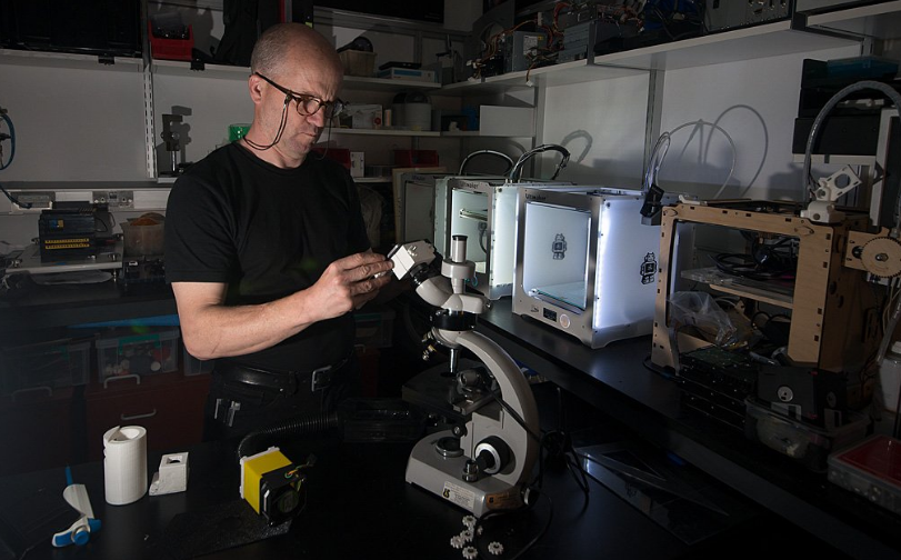
Featured image shows Professor Reynaud developing the Naiad 3D bioprinter. Photo via UCD.