Shining 3D, a Chinese 3D printing company, has announced a partnership with AGE Solutions, Italian dental software developers, to create a new orthodontics design software for the dental market.
This new software is based on AGE Solutions Maestro 3D Ortho Studio software package, which uses high-quality 3D patient-specific data to support orthodontic procedures.
“We are excited to team up with AGE Solutions,” said Oscar Meza, Vice President of Global Sales at SHINING 3D. “Our respective organizations follow the same principle of empowering the users by providing a powerful solution integrating both hardware and software, which is straightforward, cost-effective and user-friendly.”
A new digital dental solution
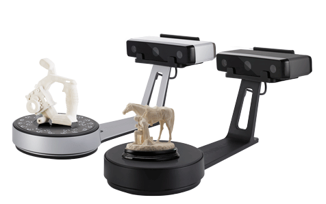
Founded in 2004, Shining 3D is the developer products including the EinScan-SP Desktop 3D Scanner, EP-M250 SLM 3D Printer, and the Geomagic Design CAD software used for medical and dental applications.
The field of orthodontics specializes in treating misaligned teeth and facial development with dental devices such as braces, headgear, and retainers.
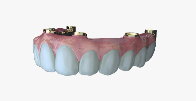
With its range of digital solutions, Shining 3D will integrate Maestro 3D Ortho Studio software, a precise CAD software using patient-specific 3D data for orthodontics-oriented inspection and editing and analysis.
The software consists of two main macro modules including the orthodontic module (Ortho Studio) and the dental restoration module (Dental CAD). In addition, Maestro Ortho Studio includes modes such as the virtual setup, brackets placement, models builder, and clear aligner, which enables the production of optimized dental devices.
Through an open standard file format, Maestro 3D Ortho Studio also provides a streamlined workflow which includes a clinic, doctor, patient, and case database. According to Andrea Spinelli, CEO of AGE Solutions, “the goal of the partnership is to make the use of 3D technologies available for the masses,” through the integration of dental digital technologies.
Shining 3D’s expands in Europe and Asia
Last year, HP announced a partnership with Shining 3D that sought to deploy over 50 HP Multi Jet Fusion 3D printers at Shining 3D facilities including Beijing, Chengdu, Guangzhou, Nanjing, and Shanghai. This Asia-Pacific expansion leveraged Shining 3D’s large clientele which includes over 10,000 worldwide.
Prior to this, 3D Printing Industry visited the opening of Shining 3D’s EMEA office in Stuttgart, Germany. During the grand opening, Shining 3D release its desktop 3D scanners, the EinScan-SE (Scan Elite) and EinScan SP for automated 3D scanning.
Keep up with the latest news in 3D printing by subscribing to the 3D Printing Industry newsletter. Also, follow us on Twitter, and like us on Facebook.
On the lookout for new talent or seeking a career change? Search and post 3D Printing Jobs for opportunities and new talent across engineering, marketing, sales and more.
Featured image shows Maestro 3D Ortho Studio software. Image via AGE Solutions.
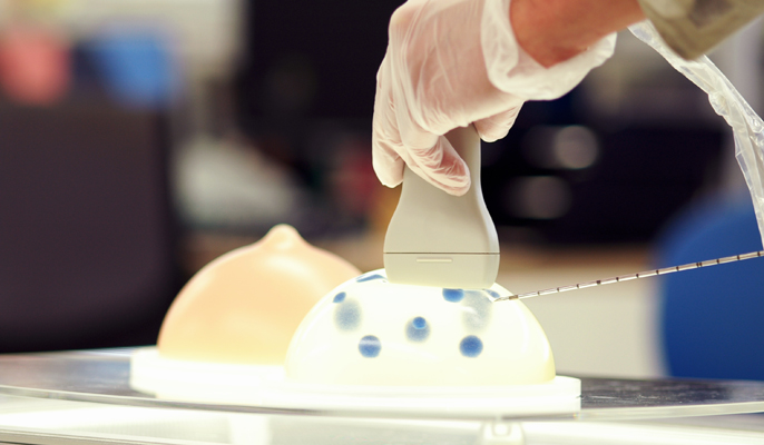
Researchers from the U.S. Food and Drug Administration (FDA) in Maryland have developed a new 3D printing software tool that aids in the creation of patient-specific 3D printed breast phantoms.
Breast phantoms are frequently used in place of breast tissue as test beds for mammography devices to ensure optimized breast cancer detection and treatment. Cancer Research UK estimates that more than 90% of women diagnosed with breast cancer at the earliest stage survive their disease for at least 5 years; this diagnosis relies on the performance capabilities of mammography systems.
According to the research published in the Journal of Medical Imaging:
“With the introduced open-source software, researchers can easily create a collection of printed phantoms that reproduce the anatomic variability of real breasts, including varying densities, heterogeneous structures, architectural distortions, and benign and malignant lesions.”
The 3D Mammoreplicator
Lead researcher of the 3D printed breast phantoms study, Andreu Badal, Physicist at the FDA suggests that although typical phantoms enable clinicians to avoid exposing patients to unnecessary mammography radiation, its predetermined shape does not accurately represent the composition of patients’ breasts.
Considering this hindrance, Badal and researchers from the University of Maryland (UMD) developed an open-source program – the mammoreplicator – which converts standard 2D mammograms into accurate 3D virtual model replicas.
The 3D models can then be fabricated into a physical phantom, demonstrated by FDA researchers with the use of three mammograms drawn from the U.S. National Institute of Health (NIH) Cancer Genome Atlas.
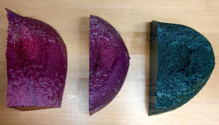
Using the Stratasys Objet260 Connex3, the research team printed one of three breast phantoms in an estimated ten hours. The 3D printed models were composed of Tango Black Plus, PolyJet photopolymers, i.e. VeroMagenta, and polymethyl methacrylate (PMMA), an acrylic glass material. In addition, each model required approximately $220 worth of raw materials.
After comparing mammograms from the 3D printed breast phantoms, the researchers found that both new and original mammograms scored similar on their structural similarity quality assessment index. The results of their research also found that the texture of the glandular and adipose tissues – found in the breast – were well-replicated in the 3D printed phantom mammograms.
3D printed breast cancer detector
Last year, the University of Twente (UT) in the Netherlands developed the 3D printed Stormram 4 robot, which is designed specifically for the collection of cells used for biopsy.
The Stormram 4, can be used directly within the magnetic chamber of an MRI scanner to provide a rate of precision impossible to achieve by hand. In addition, it is made using a high-resolution polyjet 3D printer and has been developed as a proof-of-concept for market development.
Keep up with the latest news in 3D printing by subscribing to the 3D Printing Industry newsletter. Also, follow us on Twitter, and like us on Facebook.
Looking for new talent or seeking a career change? Search and post 3D Printing Jobs for opportunities and new talent across engineering, marketing, sales and more.
Featured image shows 3D printed breast mammography phantoms. Image via Andreu Badal.
Evo Dental, a biotechnology company based in London has secured an £4 million investment from the British Growth Fund (BGF) to advance its 3D printing jaw correction clinics across the UK.
Through the new clinics, Evo Dental will provide its proprietary full jaw dental implant solution, which features digital scanning, prosthesis additive manufacturing, and high-precision milling machines to patients in need of jaw reconstruction.
“Evo is an example of the best of British craftsmanship, engineering and technology,” said Alistair Brew, Investor at BGF.
“The company has built a reputation for clinical excellence and is at the forefront of a nascent, but potentially very large market and we’re delighted to be backing them.”
One-day dental solutions
According to the NHS’ Adult Dental Health survey, more than 5 million people in UK could benefit clinically from dental implant treatments. 3D printing in particular is proving to be a growing technology for the field.
Recognizing this, Dr. R.P. Vijayanarayanan, Founder and CEO of Evo Dental, and a team of dental surgeons, dental technicians, and clinical CAD designers, developed the one-day Evo Solution treatment. This provides bespoke dental prosthetics for full jaw reconstruction surgery.
“Evo is taking what’s possible in dentistry today far beyond the limits of what has previously been available,” added Dr. Vijayanarayanan.
“We’ve combined state-of-the-art engineering, innovation and first-class patient care to deliver a solution for the chronic types of tooth problems that inflict millions of people across the UK.”
Innovation in healthcare treatments
The BGF, that previously invested £4 million in Hobs, the parent group of 3D service bureau Hobs Studio, has invested an estimated £100 million into healthcare and medical technology companies across the UK.
For Evo Dental, the BGF investment was led by Brew, Brew’s follow investor Thomas McDonnell, and George Tsangarides, a BGF Investment Manager.
Currently, Evo Dental have completed more than 3,000 full-arch reconstructions for patients.
Keep up with the latest news in 3D printing by subscribing to the 3D Printing Industry newsletter. Also, follow us on Twitter, and like us on Facebook.
On the lookout for new talent or seeking a career change? Search and post 3D Printing Jobs for opportunities and new talent across engineering, marketing, sales and more.
Featured image shows dental technicians inside the Evo Dental laboratory. Photo via Evo Dental.
Print My Part, a Cambridge-based 3D printing service, and Ramiro Joly-Mascheroni, a PHD student within the Cognitive Neuroscience Research Unit at City, University of London, have collaborated to create a range of 3D printed visual development stimulus models for prematurely born babies.
As part of Joly-Mascheroni’s research into neurocognitive mechanisms, the range of 3D printed visual models is currently being used within European hospitals to rehabilitate underdeveloped visual system within babies born before a full-term pregnancy.
3D printing a brighter future
According to the World Health Organisation (WHO), approximately 15 million babies are born prematurely every year. Within this amount, one in 20 is likely to be born blind or visually impaired – a 22% increase within the past decade.
Considering this growing dilemma, Joly-Mascheroni wanted to create an accessible and effective stimulus that trains the eye-gaze and overall sight of premature babies while inside their neonatal incubators.
Babies completely rely on neonatal incubators for survival during the first few months of life outside the womb, thus, doctors have used this time period as an opportunity to intervene in their basic visual motor skills development.
Within the neonatal units, medical professionals commonly use basic 2D images of colourful patterns, objects, animals, and emoticons to help a child focus their gaze on particular subjects while developing their retina and optic nerves.

Creating the models in a one-stop shop
A team at the Cognitive Neuroscience Research Unit approached Print My Part to help design and print the 3D visual aid models.
Using a Stratasys uPrint SE Plus 3D printer with FDM technology, the team at Print My Part was able to produce child-friendly and effective visual stimulation models.
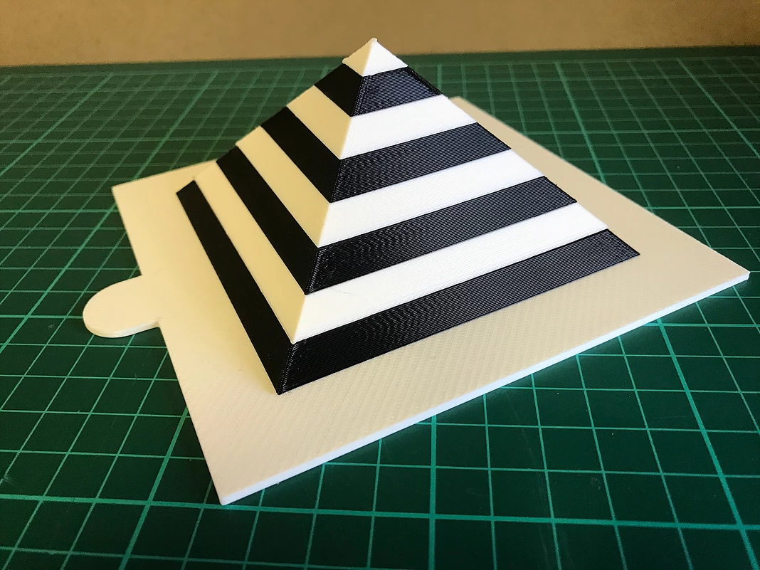
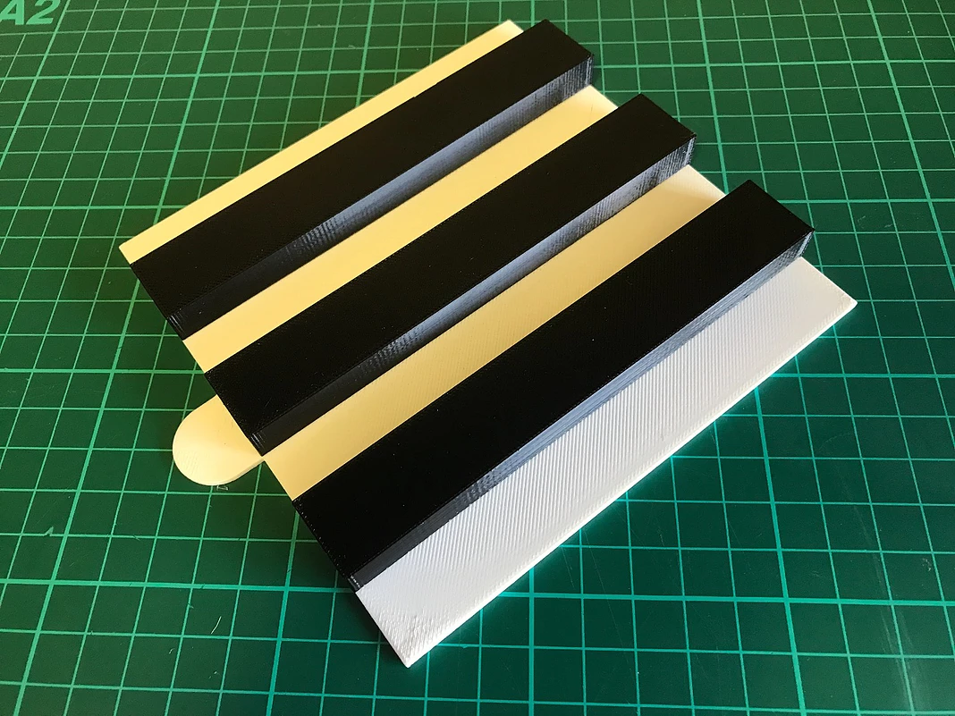
Previous medical research suggests that black and white contrasts register powerfully on a baby’s retina, sending the strongest visual signals to the baby’s brain. Therefore, Print My Part made up of a number of simple black and white slices which were assembled together to form a variety of spherical and pyramid-based shapes.
In addition, to allow better handling of the models, hold points were added to the base plates of each shape. Currently, the team is designing a mounting part to be incorporated on the models, making it easily attachable to incubator doors.
Print My Part and the Cognitive Neuroscience Research Unit teams are also considering the SLA 3D printing process to print the visual models as it complies with the EU regulations concerning sterilization.
3D printing and child health care
Additive manufacturing has demonstrated several breakthrough applications within medical treatments for children.
3D printing has been used as a surgical planning aid among surgeons across the world to ensure a safe and successful surgical operation. Recently, researchers at the Great Ormond Street Hospital (GOSH) created 3D printed replicas of children’s hearts for surgeons to better plan complex and crucial heart surgeries.
Furthermore, volunteer organization 3DP4ME are currently raising funds for a project which aims to provide 3D printed hearing-aid earmolds to disadvantaged populations in the Middle East.
Using its combination of 3D scanning and 3D printing, 3DP4ME hope to manufacture 1000s of hearing aid molds in a year.
Catch up with the latest in 3D printing by subscribing to the 3D Printing Industry newsletter. Also, follow us on Twitter, and like us on Facebook.
Looking for a change of pace or seeking new talent? Search and post 3D Printing Jobs for opportunities and new talent across engineering, marketing, sales and more.
Featured image shows 3D printed spherical visual aid model. Photo via Print My Part.
A team of researchers at the University Medical Centre (UMC) Utrecht in the Netherlands are experimenting with 3D bioprinted tissues that can be implanted into a living joint affected by arthritis. The method is hoped to allow the replacement of damaged cartilage.
Arthritis is a common disease affecting approximately 350 million people worldwide. With over 100 types of arthritis, this disease involves the disintegration of cartilage tissue found in joints, leading to pain, stiffness, and inflammation.
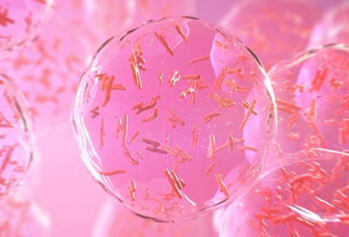
Using 3D bioprinting, patients can receive cell-specific and on-demand cartilage that would potentially grow as original cartilage to strengthen joints and reduce joint pain.
“Printing is not the last step in biofabrication, since printing something in the shape of a heart does not make it a heart,” said Jos Malda, Professor of Biofabrication in Translational Regenerative Medicine at UMC Utrecht.
“The printed construct needs time and the correct chemical and biophysical cues to mature into a functional tissue.”
Bioinks and experimentation
As part of the 3D JOINT project, funded under the EU’s Horizon 2020 vision to encourage research innovation throughout the continent, Professor Malda and his team are building upon the capabilities of 3D printers and the deposition of stem cells.
Using the 3D bioprinting technique and following a precise medical blueprint, stem cells are deposited by 3D printers creating complex tissues layer by layer.
As demonstrated by Newcastle University’s 3D bioprinted human corneas, bioink solutions created from donor stem cells and hydrogels – a material class that consists of large molecules such as polymers – can produce living conditions for organisms within the human body.
With the challenge of maintaining the right conditions for a cellular building material, Professor Malda integrated hydrogel into cartilage tissue.
“For bioprinting, the material has to be able to keep cells alive. This demands aqueous conditions and processing under a relatively low temperature, which makes hydrogel-based materials ideal candidates.”
Yet, the soft nature of such hydrogels comes at a disadvantage, according to Professor Malda. This is due to its inability to withstand the mechanical forces certain tissues undergo in the body.
Onwards to additive materials
As a result of their findings, the UMC Utrecht research team began experimenting with additive materials that can strengthen hydrogels in order for them to act as replacement cartilage.
“Reinforcing the hydrogel makes it stronger – just like steel rods are combined with soft cement to create the reinforced concrete that makes the foundations of our homes,” said Professor Malda.
His team is using a 3D printing technique called melt electro-writing that combines melted polycaprolactone, a type of polyester, with an electrical field that creates fibres as thin as a hair to create scaffolding.
“The combination of the hydrogel with the fibres acts in synergy, increasing the strength of the composite over 50 times while still allowing the cells to generate extracellular matrix and mature into a cartilage-like tissue.”
His team is now developing this process to create larger structures while including different materials for combined bone and cartilage tissue replacements. This will ultimately reach UMC Utrecht’s goal to eventually 3D print a complete joint.
Also using the 3D bioprinting technology is BIOLIFE4D, a biotechnology company from Chicago, who successfully demonstrated the ability to 3D print human heart tissue.
Keep up with the latest in 3D printing by subscribing to the 3D Printing Industry newsletter. Also, follow us on Twitter, and like us on Facebook.
Looking for a change of pace or seeking new talent? Search and post 3D Printing Jobs for opportunities and new talent across engineering, marketing, sales and more.
Featured image shows an x-ray of arthritic hands. Image via Medical Xpress.
According to the British Heart Foundation (BHF), each year, there are over 4600 babies born with Congenital Heart Disease (CHD) – potentially life-threatening defects within the structure of the heart.
With the unique qualities of each defect, researchers at the Great Ormond Street Hospital (GOSH) in London are developing 3D printed replicas of children’s hearts for surgeons to better plan complex and crucial heart surgeries.
According to Dr. Claudio Capelli, a BHF-funded researcher leading the 3D printed hearts project at GOSH, “This can mean quicker, more effective surgeries, which can be an advantage for the patients, as they can recover faster with fewer complications.”
The case of Lucas Ciulean
Dr. Capelli and his team of researchers are explored surgical solutions for children with CHD through life-size 3D models of the patient’s heart. He explains, “Having a 3D printed heart made can also help adults, but it is particularly helpful for children with congenital heart disease as their hearts are very small and particularly complex.”
In one case, Dr. Capelli’s work has contributed to the successful surgery of Lucas Ciulean, a 13-month-old born with a rare heart defect. Lucas’ aorta, the main artery distributing blood around the body, was connected to the wrong part of his heart.
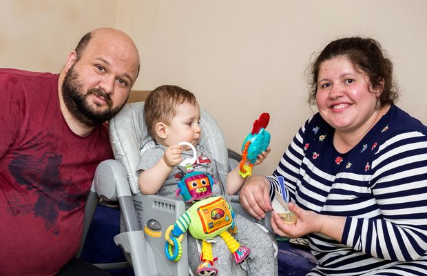
Using a medical scan to recreate a 3D model of Lucas’ heart helped surgeons understood how the aorta was positioned with respect to the pulmonary artery – which transports blood from the heart to the lungs.
Following their son’s successful surgery and recovery, Lucas’ parents expressed their admiration for the 3D printed models. His father, Tiberius Ciulean, said, “When the doctor first showed us the heart, we were amazed, we had no idea about this technology and that they were printing 3D hearts,”
“To have Lucas’ heart in your hand and hear everything that the surgeon did and about the problem, it was amazing.”
Heart models in hours
Creating the 3D model of Lucas’ heart began with combining a series of medical scans. The scans are sent and loaded into 3D printing software where technicians orientated the heart and choose the material and surface finishes.
Using a resin-based 3D printer, the research team printed layers as small as 32 microns (0.032mm) to form the shape of the heart and then used a UV lamp to solidify the model.
According to Dr. Capelli, 3D printing a baby’s heart requires up to 2000 layers and three to four hours to build, while an adult’s heart can take up to 10,00 layers and must be fabricated overnight.
After 3D printing, the model of Lucas’ heart was then pressure washed and cleaned to remove excess resin. After this process, the 3D model was sent to the surgeon and cardiologist associated with the surgery.
3D printed organs & surgeries
Last year, the Queen Elizabeth Hospital in Birmingham, U.K. reduced surgical planning by an estimated 93% through the use of 3D models for maxillofacial (face and jaw) surgeries.
Also contributing to the research within children cardiology is the Children’s Heart Research and Outcomes Center (HeRO) who are developing 3D bioprinted valves, leaflets, and patches to provide a long-lasting solution for children born with CHD.
Dr. Capelli is now working on BHF-funded research to understand the effects of patient’s seeing and touching their own 3D-printed heart and whether that helps teenagers better cope with their heart condition.
Keep up with the latest innovations in 3D printing materials by subscribing to the 3D Printing Industry newsletter. Also, follow us on Twitter, and like us on Facebook.
Looking for a change of pace or seeking new talent? Search and post 3D Printing Jobs for opportunities and new talent across engineering, marketing, sales and more.
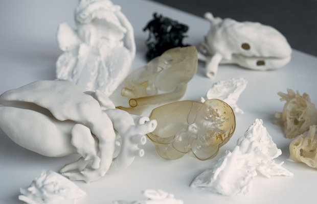
Featured image show 3D models of hearts printed by researchers from the British Heart Foundation. Photo via the British Heart Foundation.
Engineering students from the University of Alabama in Huntsville (UAH) have 3D printed medical training tools to be used by undergraduates in UAH’s College of Nursing. With the creation of 3D devices such as a cricothyrotomy trainer, vein finder, and an onychectomy trainer, aspiring nurses can safely practice important medical procedures on lifelike healthcare simulators.
Making medical task tools
Recognising the growing trend of 3D printed medical models used for task training, Dr. Lori Lioce, Clinical Associate Professor at UAH ‘s College of Nursing and Norven Goddard, a Research Scientist at UAH’s Systems Management and Production (SMAP) Center decided to use the university’s 3D printers create their own models.
“These models cost more than a thousand dollars, but we wanted something that would save money, be cost-effective and use the university’s resources,” said Goddard. “We asked ourselves, how cheaply can we do this?”
Lioce enlisted a “3D specialist team” comprising of engineering and computer science students to help produce the medical task trainers. Firstly, the team created a cricothyrotomy trainer – a training model of a patient’s neck, used to practice emergency procedures to clear a patient’s airway. The task trainer’s digital design files are available on the 3D file sharing platform, Thingiverse, and after three prototypes, the team created over four accessible models for all medical classes.
Next, the team 3D printed an onychectomy trainer, a device used to practice the removal of thumbnails as well as a 3D printed vein finder which, according to Goddard, typically cost hundreds of dollars. Nonetheless, the student team was able to create these devices using $6 open-source design files.
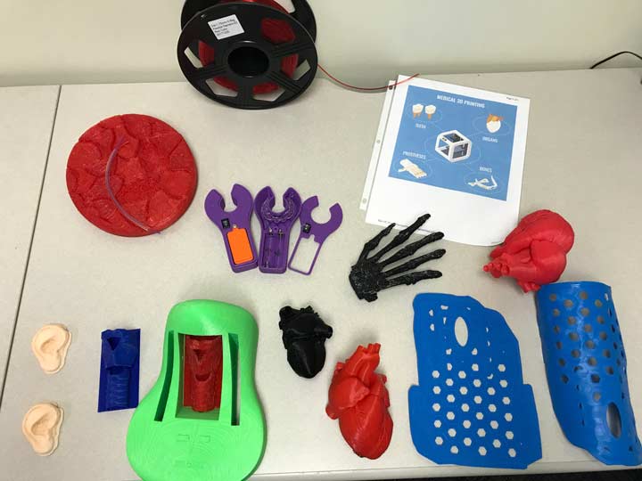
With the use of 3D printing, the College of Nursing has saved an estimated $6000 on medical training equipment and has given nurse practitioner students at UAH the opportunity to repeatedly practice a specific skill in preparation for providing healthcare in the real world.
After the success of the 3D printed healthcare simulators, Lioce, Goddard and the student team plan to teach other nursing students how to 3D print with upcoming projects to create an injection simulation pad and 3D medical models.
“We’re trying to cross-pollinate so everyone knows how to 3D print, injection mold, solder, use the software and do whatever else is needed,” said Goddard.
Additive Manufacturing provides realistic healthcare simulations
The customizable capabilities of additive manufacturing technologies, as well as the plethora of unique 3D printing material, have paved the way for more realistic healthcare simulation devices for medical students.
Researchers from the Japanese Tottori University Hospital previously collaborated with medical company Tmsuk R&D Inc to create a medical simulation robot with 3D printed organs. This robot has been designed to exhibit human emotions and reflexes to give medical professionals and students gain a greater awareness of the patient experience.
Furthermore, 3D Systems has previously launched the medical training tool, the Simbionix SPINE Mentor, which combines virtual reality simulation, 3D printing and medical tools to create a hybrid solution that medical practitioners train for spinal surgery training
Catch up with the latest in additive manufacturing by subscribing to the 3D Printing Industry newsletter. Also, follow us on Twitter, and like us on Facebook.
Looking for a change of pace or seeking new talent? Search and post 3D Printing Jobs for opportunities and new talent across engineering, marketing, sales and more.
Featured image shows SMAP Center student interns James Tovar, Marquis Myler, Nicholas Swinford, Martavia Lucious, Matthew Daigle, and Andrew Farris who created a variety of cost-effective 3-D printed task trainers to be used by students in UAH’s College of Nursing.
Additive manufacturing continues to aid in the production of medical-grade implants. From 3D printed cages built to support the spine to the development of biocompatible facial implants, 3D printed implants have displayed its functionalities over traditional medical procedures.
Taking a step further to improve spinal treatments, Centinel Spine, a Pennsylvania-based spinal device development company, has received 510(k) clearance from the U.S Food and Drug Administration (FDA) to market its 3D printed implantation devices.
Centinel Spine’s FLX Platform consists of a series of 3D printed all-titanium fusion devices that will work to stabilize vertebrae from the front (anterior) of the spine in order to increase the healing process for patients after spinal surgeries.
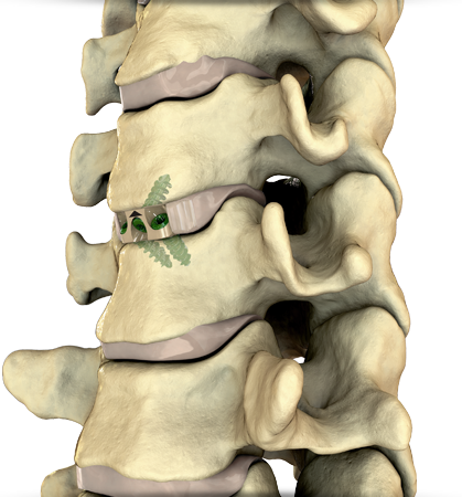
The FLX devices
Centinel Spine was founded through a merger-acquisition between surgical devices manufacturers, Raymedica and Surgicraft and has continued to lead the development of anterior spine technology with its latest development, the FLX devices.
These devices are made from a combination of solid and porous sections which are transparent to x-rays (radiolucent) – erasing the chance for image distortion. As a result of this composite material, the implant’s visibility during surgery is increased as well as its flexibility when compared to the stiffness of solid titanium implants.
Centile Spine’s innovative technology, STALIF (Stand Alone Lumbar Interbody Fusion), also enables its implants to have a biocompatible structure which permits tissue ingrowth within bones concerning the spine. This encourages a quicker fusion between the bone and implant, contributing to spinal support.
“The clearance of the FLX Platform represents the next evolution in STALIF technology,” said John Viscogliosi, CEO and Chairman of Centinel Spine. “Utilizing 3D-printing, we are able to offer the proven benefits of the STALIF design in a truly novel, all-titanium lattice option,”
“This allows our surgeons the flexibility to use multiple implant material options through a single set of instruments to address each patient’s unique pathology.”
An increase in AM medical devices
Over the past few years, 3D printed devices within the healthcare sector have significantly increased. According to FDA Commissioner Scott Gottlieb, M.D, the FDA has reviewed and cleared over 100 3D printed medical devices that are currently on the market. This includes K2M, a medical device company based in Virginia, and its 3D printed spinal implant which can now be used inside the human body to help correct spinal defects as well as EIT’s 3D printed titanium cellular implants.
Centinel Spine believes that the clearance of the FLX platform is the next step in its mission to become the worldwide leading spine company with the widest platform breadth and depth of technology.
Keep up with the latest advancements in additive manufacturing as they happen. Subscribe to the 3D Printing Industry newsletter, follow us on Twitter, and like us on Facebook.
Looking for a change of pace or seeking new talent? Search and post 3D Printing Jobs for opportunities and new talent across engineering, marketing, sales and more.
Featured image shows close-up graphic of the 3D printed all-titanium implant device. Image via Centinel Spine.
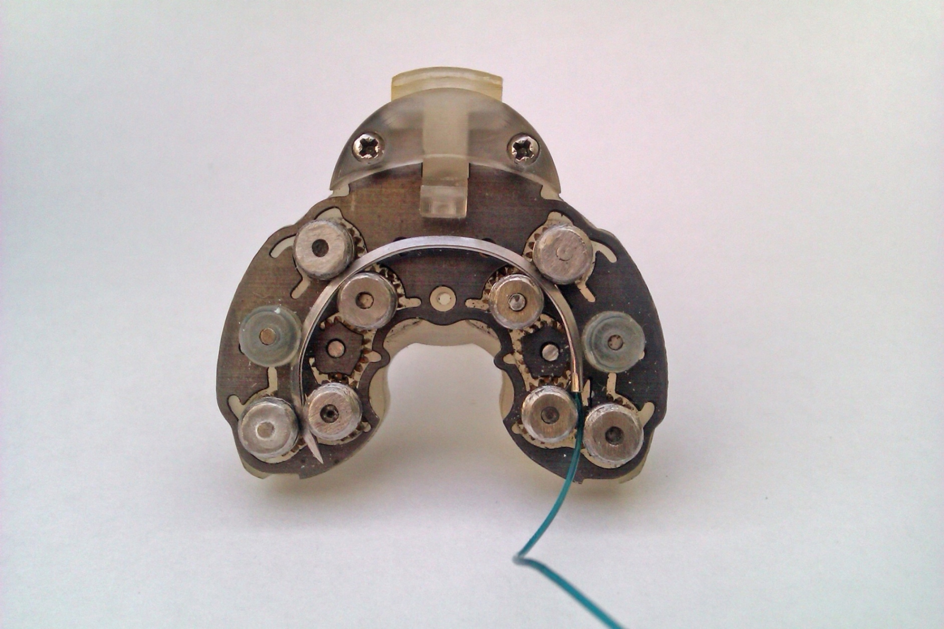
Sutrue, a UK-based medical technology company, has debuted its new 3D printed automated suturing devices at The Design Museum, London. 3D Printing Industry attended the launch to learn more about how additive manufacturing is shaping the future of medicine.
Medical stitching (suturing) has been done by hand for over 5000 years, causing numerous needlestick injuries and diseases and time-consuming procedures. Sutrue hopes to address these issues with its two new innovations; the handheld and robotic automated suturing devices. The medical devices increase suturing accuracy, reduce the risk of injuries and diseases, and could potentially save the NHS an estimated £10.7 million each year.
Alexander Berry, Sutrue founder, and Director explained that an intricate metal 3D printed gear mechanism operates the needle of both devices.
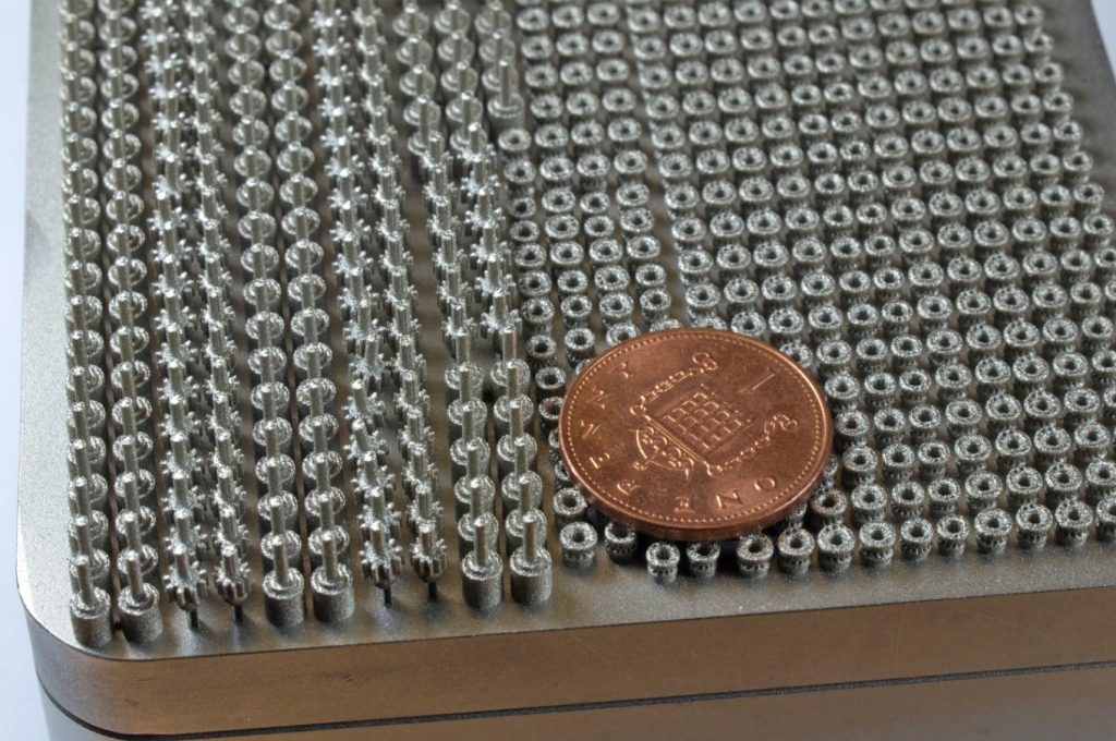
GE Additive streamlines production
The metal gears in the Suture devices are 250 microns in length, while the tooth of an individual gear is 400 microns, boasting a higher level of accuracy within production. The medical devices were 3D printed by ES Technology using Concept Laser MLab Cusing additive manufacturing systems.
Berry explained to guests at the Design Museum that, “Without the help from GE and Concept Laser, we would have spent years going back and forth during prototype development. That’s the advantage of 3D printing – its speculative which allows us to fail during the development of our products.”
The Sutrue founder then went on to emphasize that using a MLab Cusing metal 3D printer during the engineering process allowed rapid production of 38 prototypes for the automated suturing devices.
Stephan Zeidler, Business Development Manager in the medical sector at Concept Laser told me, “We were happy to help Sutrue on their journey to improve healthcare. 3D printing is already established with many medical companies so this was nothing new for us.”
The automated handheld suturing device
The Sutrue team worked with cardiologist Dr. Richard Trimlett to help translate the manual stitching specifications into the handheld device. The miniature gears allow a rotating needle to tilt and adjust to suit the angle of a surgeon during cardiac, thoracic, cranial and gastrointestinal operations.
I had the opportunity to use the device, it was comfortable to hold and fairly lightweight. I pressed one of the two buttons which activated the needle immediately. However, an inexperienced user (such as myself) found it hard to complete a full stitch.
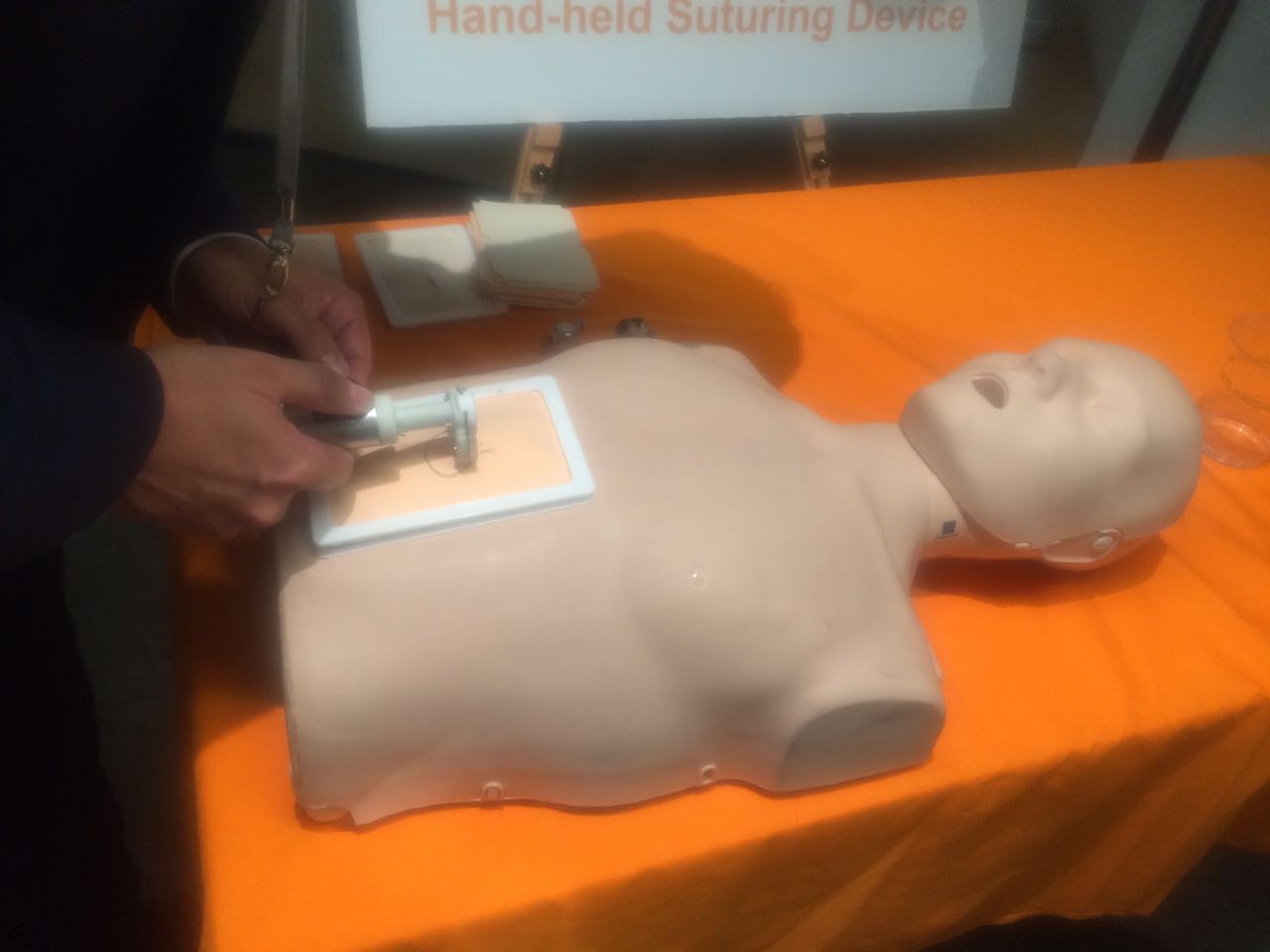
For medical professionals, the handheld automated suturing device creates a smoother and faster stitch when compared to traditional methods. The device is able to stitch minimally invasive keyhole incisions. This reduces the need for longer and deeper high-risk cuts that are more difficult to close when stitching.
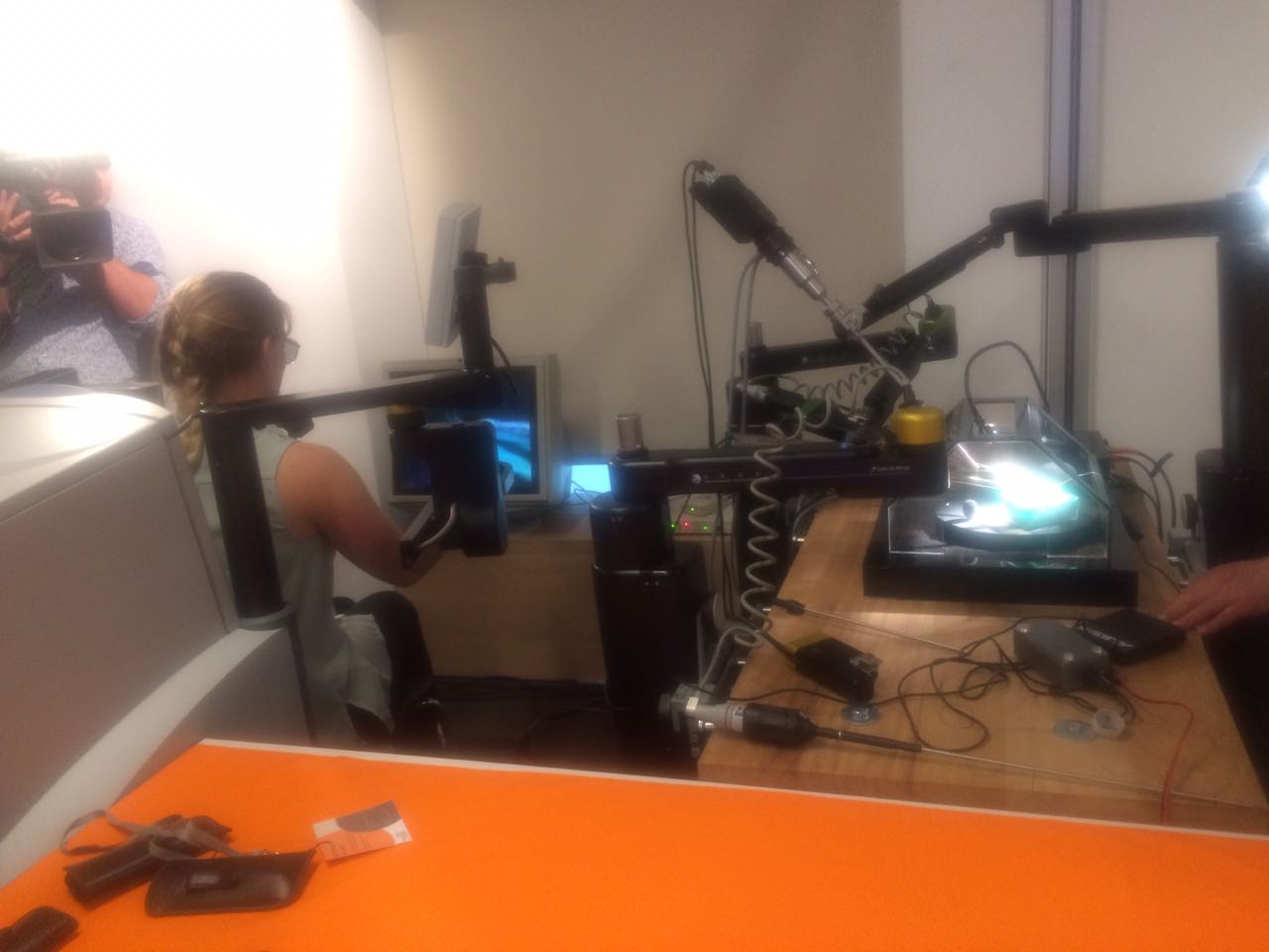
The robotic automated device
During the Q and A portion of the press event, the Sutrue team discussed that the robotic automated device came after the handheld device. Once the handheld device was completed, the engineers were able to apply the angles from a surgeon’s perspective to the Zeus medical robot.
To test the robotic device at the press event, Berry was required to wear a pair of 3D glasses. This enabled him to view the separate computer monitor where the suturing activity was taking place.
Berry demonstrated the speed of the device by producing a single stitch in under a second, as opposed to an experienced surgeon, who can take up to 25 seconds.
Dr Trimlett believes that this device can be influential for the future of robotic surgery due to its ability to perform more low-risk keyhole surgeries at a faster rate.
The Queen Elizabeth Hospital in Birmingham, UK have also recently implemented 3D Printing technology to improve surgical time with the Stratasys Objet Eden350V 3D Printer.
Future commercialization for devices
Sutrue plans on commercializing their automated devices within hospitals and veterinary and dental practices. Sutrue has also been approached by bespoke clothing manufacturers with the potential to use their automated suturing devices for fabric stitching.
For more of the latest 3D printing product developments subscribe to the 3D Printing Industry newsletter, follow us on Twitter and like us on Facebook.
Looking for a change of pace? Sign up for 3D Printing Jobs here.
Featured image shows Additively manufactured parts of the automated suturing device on the build plate of an Mlab cusing from Concept Laser. Photo via Concept Laser